Review
. 2012 Mar;132(3 Pt 2):785-96.
doi: 10.1038/jid.2011.426. Epub 2012 Jan 5.

Shining a light on xeroderma pigmentosum

Affiliations
Review

Shining a light on xeroderma pigmentosum

John J DiGiovanna et al. J Invest Dermatol. 2012 Mar.

Abstract

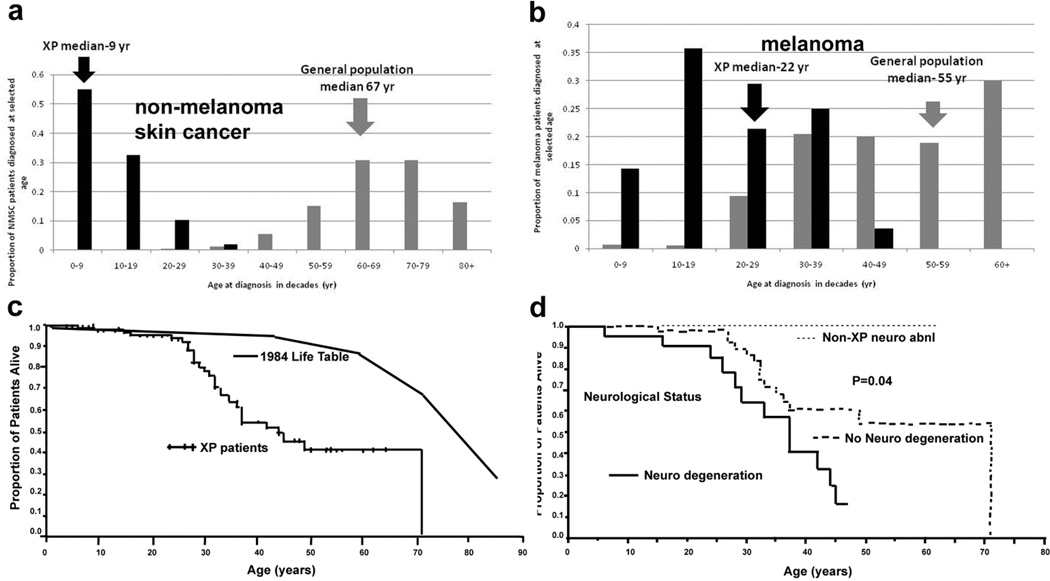
Xeroderma pigmentosum (XP) is a rare, autosomal recessive disorder of DNA repair characterized by sun sensitivity and UV radiation-induced skin and mucous membrane cancers. Initially described in 1874 by Moriz Kaposi in Vienna, nearly 100 years later, James Cleaver in San Francisco reported defective DNA repair in XP cells. This eventually provided the basis for a mechanistic link between sun exposure, DNA damage, somatic mutations, and skin cancer. XP cells were found to have defects in seven of the proteins of the nucleotide excision repair pathway and in DNA polymerase η. XP cells are hypersensitive to killing by UV radiation, and XP cancers have characteristic "UV signature" mutations. Clinical studies at the National Institutes of Health found a nearly 10,000-fold increase in skin cancer in XP patients under the age of 20 years, demonstrating the substantial importance of DNA repair in cancer prevention in the general population. Approximately 25% of XP patients have progressive neurological degeneration with progressive loss of neurons, probably from DNA damage induced by oxidative metabolism, which kills nondividing cells in the nervous system. Interestingly, patients with another disorder, trichothiodystrophy, have defects in some of the same genes as XP, but they have primary developmental abnormalities without an increase in skin cancer.


Conflict of interest statement

CONFLICT OF INTEREST

The authors declare no conflict of interest.

Figures

Figure 1
Figure 1
XP and TTD patients studied. a) Patient XP420BE complementation group XP-D at 9 months of age with severe blistering erythema of the malar area following minimal sun exposure. Note sparing of her forehead and eyes that were protected by a hat. b) Patient XP358BE (XP-C) at age 2 years did not sunburn easily but developed multiple hyperpigmented macules on her face. A rapidly growing SCC or keratoacanthoma grew on her upper lip and a pre-cancerous lesion appeared on her forehead. c) Northern African patient XP393BE (XP-C) (Mahindra et al. 2008) at age 23 years with numerous hyperpigmented macules on his face. Nodular basal cell cancer is present on his left nasal root. Pigmented basal cell cancer is present on his left cheek. His eyes show cornea scarring from unprotected sun exposure. d) Patient XP19BE (XP-A) (Robbins et al. 1991) at age 35 years with neurological degeneration. He has numerous hyperpigmented macules on sun exposed areas of his face and neck. Progressive sensorineural deafness requires use of a hearing aid. Images a–d from (Bradford et al. 2011) e. Corneal clouding, pterygium, contact lens, loss of lashes on lower eyelid. f. Sharp demarcation between the poikilodermatous changes seen in sun exposed skin compared to double covered area of the buttocks of 35 year old XP patient. g. Loss of vermillion border of the lips with prominent telangiectasias and scaring of the lips and anterior tongue. h: squamous cell carcinoma of the anterior tongue in African man (from (Mahindra et al. 2008)). i–j Clinical appearance of TTD. i : 3 yo girl with short brittle hair which is sparse and broken off at different lengths. She rarely has haircuts except to trim uneven areas. She has a smiling, outgoing personality typical of TTD. j. Tiger tail banding under polarizing microscopy (Original magnification × 10.) (Images i–j from (Liang et al. 2005).
Figure 2
Figure 2
XP skin cancer by age at first skin cancer diagnosis and skin cancer type and mortality compared to U.S. general population. a. Proportion of NMSC patients diagnosed at selected ages. b. Proportion of melanoma patients diagnosed at selected ages. Individuals with both NMSC and melanoma were used for both analyses. General population data taken from (Glass and Hoover 1989). c. Kaplan Meier curve of xeroderma pigmentosum patient survival compared to US general population: 30% of XP patients had died by age 32. The survival of the XP patients was significantly less than the general population (p<0.001). d. Kaplan Meier curve of xeroderma pigmentosum patient survival stratified by neurologic phenotype. Patients with neurologic degeneration had poorer survival rates than those without neurologic degeneration (p=0.04). (Graphs from (Bradford et al. 2011)).
Figure 3
Figure 3
Nucleotide excision repair (NER) pathway. Transcription coupled repair (TCR) removes damage from actively transcribing genes while global genome repair (GGR) removes damage from the remainder of the genome. In GGR damage such as ultraviolet induced cyclobutane pyrimidine dimers (CPD) or 6-4 photoproducts (6-4 PP) are recognized by proteins including the XPE (DDB2) and XPC gene products. In TCR, the lesion appears to block the progress of RNA polymerase II in a process involving the CSA and CSB gene products. Following initial damage recognition the pathways converge. The XPB (ERCC3) and XPD (ERCC2) helicases unwind the region surrounding the lesion along with the XPA and XPG (ERCC5) gene products, and replication protein A (RPA). The XPF and XPG (ERCC5) endonucleases perform incisions to remove the lesion in a fragment of about 30 nucleotides. The resulting gap is filled in by de novo DNA synthesis. This system is coordinated so that if one part of the pathway is mutated the entire pathway fails to function normally. Mutations in the genes in rectangles have been associated with clinical disease. This diagram is modified from (Van Steeg and Kraemer 1999; Kraemer et al. 2007).
Figure 4
Figure 4
DNA repair diseases - relationship of clinical disorders (red rectangles) to molecular defects (gray ovals) in DNA repair diseases. Ten clinical diseases and 13 molecular defects are represented. One disease may be caused by mutations in several different genes. Conversely, different mutations in one gene may result in several different clinical diseases. Modified from (Kraemer 2004; Kraemer et al. 2007).

References

    1. Alekseev S, Kool H, Rebel H, Fousteri M, Moser J, Backendorf C, et al. Enhanced DDB2 expression protects mice from carcinogenic effects of chronic UV-B irradiation. Cancer Res. 2005;65(22):10298–10306. - PubMed
    1. Arase S, Kozuka T, Tanaka K, Ikenaga M, Takebe H. A sixth complementation group in xeroderma pigmentosum. Mutat.Res. 1979;59:143–146. - PubMed
    1. Blankenburg S, Konig IR, Moessner R, Laspe P, Thoms KM, Krueger U, et al. Assessment of 3 xeroderma pigmentosum group C gene polymorphisms and risk of cutaneous melanoma: a case-control study. Carcinogenesis. 2005 - PubMed
    1. Bradford PT, Goldstein AM, Tamura D, Khan SG, Ueda T, Boyle J, et al. Cancer and neurologic degeneration in xeroderma pigmentosum: long term follow-up characterises the role of DNA repair. J.Med.Genet. 2011;48(3):168–176. - PMC - PubMed
    1. Bredberg A, Kraemer KH, Seidman MM. Restricted ultraviolet mutational spectrum in a shuttle vector propagated in xeroderma pigmentosum cells. Proc.Natl.Acad.Sci.U.S.A. 1986;83(21):8273–8277. - PMC - PubMed

Publication types

MeSH terms