

Hepatitis B virus X protein inhibits extracellular IFN-α-mediated signal transduction by downregulation of type I IFN receptor
- PMID: 22218495
- PMCID: PMC3577137
- DOI: 10.3892/ijmm.2012.879
Hepatitis B virus X protein inhibits extracellular IFN-α-mediated signal transduction by downregulation of type I IFN receptor
Abstract
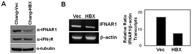
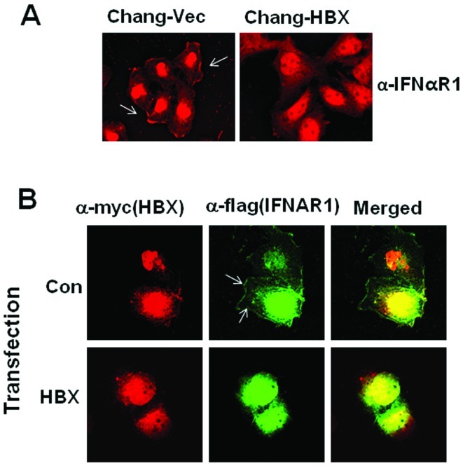
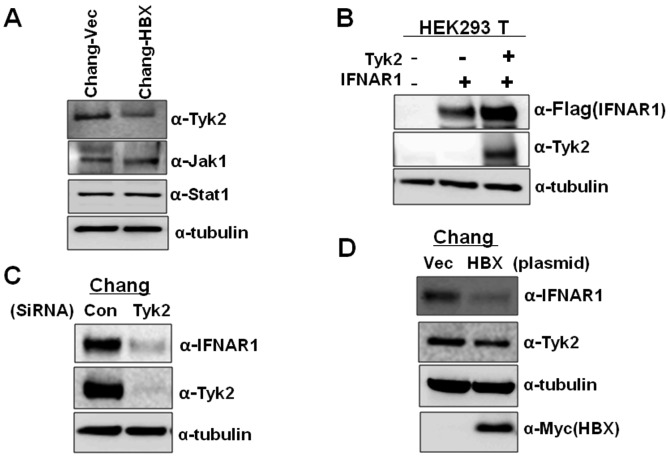
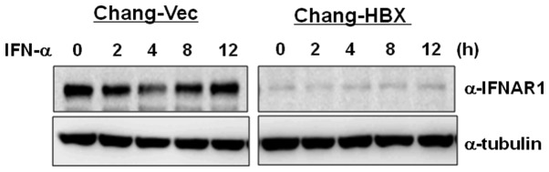
We have previously shown that hepatitis B virus (HBV) protein X (HBX), a regulatory protein of HBV, activates Stat1, leading to type I interferon (IFN) production. Type I IFN secreted from HBX-expressing hepatic cells enforces antiviral signals through its binding to the cognate type I IFN receptor. We therefore investigated how cells handle this detrimental situation. Interestingly, compared to Chang cells stably expressing an empty vector (Chang-Vec), Chang cells stably expressing HBX (Chang-HBX) showed lower levels of IFN-α receptor 1 (IFNAR1) protein, a subunit of type I IFN receptor. The levels of IFNAR1 transcripts detected in Chang-HBX cells were lower than the levels in Chang-Vec cells, indicating that HBX regulates IFNAR1 at the transcriptional level. Moreover, we observed that HBX induced the translocation of IFNAR1 to the cytoplasm. Consistent with these observations, HBX also downregulated Tyk2, which is required for the stable expression of IFNAR1 on the cell surface. Eventually, Chang-HBX cells consistently maintained a lower level of IFNAR1 expression and displayed no proper response to IFN-α, while Chang-Vec cells exhibited a proper response to IFN-α treatment. Taken together, we propose that HBX downregulates IFNAR1, leading to the avoidance of extracellular IFN-α signal transduction.
Figures
Similar articles
-
Expression of HBX, an oncoprotein of hepatitis B virus, blocks reoviral oncolysis of hepatocellular carcinoma cells.Cancer Gene Ther. 2009 May;16(5):453-61. doi: 10.1038/cgt.2008.95. Epub 2008 Dec 19. Cancer Gene Ther. 2009. PMID: 19096445
-
Interferon-gamma inhibits interferon-alpha signalling in hepatic cells: evidence for the involvement of STAT1 induction and hyperexpression of STAT1 in chronic hepatitis C.Biochem J. 2004 Apr 1;379(Pt 1):199-208. doi: 10.1042/BJ20031495. Biochem J. 2004. PMID: 14690454 Free PMC article.
-
Interferon-alpha restrains growth and invasive potential of hepatocellular carcinoma induced by hepatitis B virus X protein.World J Gastroenterol. 2008 Sep 28;14(36):5564-9; discussion 5568. doi: 10.3748/wjg.14.5564. World J Gastroenterol. 2008. PMID: 18810776 Free PMC article.
-
Identification of TRIM14 as a Type I IFN-Stimulated Gene Controlling Hepatitis B Virus Replication by Targeting HBx.Front Immunol. 2018 Aug 13;9:1872. doi: 10.3389/fimmu.2018.01872. eCollection 2018. Front Immunol. 2018. PMID: 30150992 Free PMC article. Clinical Trial.
-
The molecular basis for differential type I interferon signaling.J Biol Chem. 2017 May 5;292(18):7285-7294. doi: 10.1074/jbc.R116.774562. Epub 2017 Mar 13. J Biol Chem. 2017. PMID: 28289098 Free PMC article. Review.
Cited by
-
Hidden liver-joint axis: HBV infection causes rheumatoid arthritis via TRAFD1 with imbalance of HBV X protein and trans-ferulic acid.Virulence. 2024 Dec;15(1):2422540. doi: 10.1080/21505594.2024.2422540. Epub 2024 Nov 7. Virulence. 2024. PMID: 39484999 Free PMC article.
-
Identification of novel susceptibility loci associated with hepatitis B surface antigen seroclearance in chronic hepatitis B.PLoS One. 2018 Jul 5;13(7):e0199094. doi: 10.1371/journal.pone.0199094. eCollection 2018. PLoS One. 2018. PMID: 29975729 Free PMC article.
-
Casein Kinase 1α Mediates the Degradation of Receptors for Type I and Type II Interferons Caused by Hemagglutinin of Influenza A Virus.J Virol. 2018 Mar 14;92(7):e00006-18. doi: 10.1128/JVI.00006-18. Print 2018 Apr 1. J Virol. 2018. PMID: 29343571 Free PMC article.
-
Ubiquitination-mediated regulation of interferon responses.Growth Factors. 2012 Jun;30(3):141-8. doi: 10.3109/08977194.2012.669382. Epub 2012 Mar 7. Growth Factors. 2012. PMID: 22394219 Free PMC article. Review.
-
Intracellular interferon signalling pathways as potential regulators of covalently closed circular DNA in the treatment of chronic hepatitis B.World J Gastroenterol. 2021 Apr 14;27(14):1369-1391. doi: 10.3748/wjg.v27.i14.1369. World J Gastroenterol. 2021. PMID: 33911462 Free PMC article. Review.
References
-
- Payelle-Brogard B, Pellegrini S. Biochemical monitoring of the early endocytic traffic of the type I interferon receptor. J Interferon Cytokine Res. 2010;30:89–98. - PubMed
-
- Stark GR, Kerr IM, Williams BR, Silverman RH, Schreiber RD. How cells respond to interferons. Annu Rev Biochem. 1998;67:227–264. - PubMed
-
- Bonifacino JS, Traub LM. Signals for sorting of transmembrane proteins to endosomes and lysosomes. Annu Rev Biochem. 2003;72:395–447. - PubMed
Publication types
MeSH terms
Substances
Grants and funding
LinkOut - more resources
Full Text Sources
Other Literature Sources
Research Materials
Miscellaneous
