

Canine models of Duchenne muscular dystrophy and their use in therapeutic strategies
- PMID: 22218699
- PMCID: PMC3911884
- DOI: 10.1007/s00335-011-9382-y
Canine models of Duchenne muscular dystrophy and their use in therapeutic strategies
Abstract
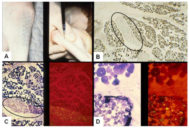
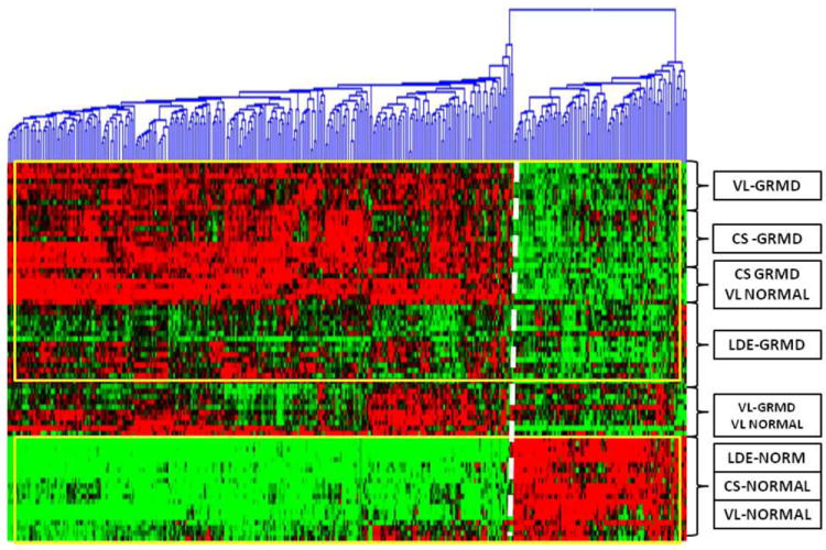
Duchenne muscular dystrophy (DMD) is an X-linked recessive disorder in which the loss of dystrophin causes progressive degeneration of skeletal and cardiac muscle. Potential therapies that carry substantial risk, such as gene- and cell-based approaches, must first be tested in animal models, notably the mdx mouse and several dystrophin-deficient breeds of dogs, including golden retriever muscular dystrophy (GRMD). Affected dogs have a more severe phenotype, in keeping with that of DMD, so may better predict disease pathogenesis and treatment efficacy. Various phenotypic tests have been developed to characterize disease progression in the GRMD model. These biomarkers range from measures of strength and joint contractures to magnetic resonance imaging. Some of these tests are routinely used in clinical veterinary practice, while others require specialized equipment and expertise. By comparing serial measurements from treated and untreated groups, one can document improvement or delayed progression of disease. Potential treatments for DMD may be broadly categorized as molecular, cellular, or pharmacologic. The GRMD model has increasingly been used to assess efficacy of a range of these therapies. A number of these studies have provided largely general proof-of-concept for the treatment under study. Others have demonstrated efficacy using the biomarkers discussed. Importantly, just as symptoms in DMD vary among patients, GRMD dogs display remarkable phenotypic variation. Though confounding statistical analysis in preclinical trials, this variation offers insight regarding the role that modifier genes play in disease pathogenesis. By correlating functional and mRNA profiling results, gene targets for therapy development can be identified.
Figures
Similar articles
-
The golden retriever model of Duchenne muscular dystrophy.Skelet Muscle. 2017 May 19;7(1):9. doi: 10.1186/s13395-017-0124-z. Skelet Muscle. 2017. PMID: 28526070 Free PMC article. Review.
-
Golden retriever muscular dystrophy (GRMD): Developing and maintaining a colony and physiological functional measurements.Methods Mol Biol. 2011;709:105-23. doi: 10.1007/978-1-61737-982-6_7. Methods Mol Biol. 2011. PMID: 21194024
-
Repression of phosphatidylinositol transfer protein α ameliorates the pathology of Duchenne muscular dystrophy.Proc Natl Acad Sci U S A. 2017 Jun 6;114(23):6080-6085. doi: 10.1073/pnas.1703556114. Epub 2017 May 22. Proc Natl Acad Sci U S A. 2017. PMID: 28533404 Free PMC article.
-
Identification in GRMD dog muscle of critical miRNAs involved in pathophysiology and effects associated with MuStem cell transplantation.BMC Musculoskelet Disord. 2016 May 11;17:209. doi: 10.1186/s12891-016-1060-5. BMC Musculoskelet Disord. 2016. PMID: 27170302 Free PMC article.
-
The value of mammalian models for duchenne muscular dystrophy in developing therapeutic strategies.Curr Top Dev Biol. 2008;84:431-53. doi: 10.1016/S0070-2153(08)00609-1. Curr Top Dev Biol. 2008. PMID: 19186250 Review.
Cited by
-
Tadalafil Treatment Delays the Onset of Cardiomyopathy in Dystrophin-Deficient Hearts.J Am Heart Assoc. 2016 Aug 9;5(8):e003911. doi: 10.1161/JAHA.116.003911. J Am Heart Assoc. 2016. PMID: 27506543 Free PMC article.
-
Dynamic changes to lipid mediators support transitions among macrophage subtypes during muscle regeneration.Nat Immunol. 2019 May;20(5):626-636. doi: 10.1038/s41590-019-0356-7. Epub 2019 Apr 1. Nat Immunol. 2019. PMID: 30936495 Free PMC article.
-
Challenges associated with homologous directed repair using CRISPR-Cas9 and TALEN to edit the DMD genetic mutation in canine Duchenne muscular dystrophy.PLoS One. 2020 Jan 21;15(1):e0228072. doi: 10.1371/journal.pone.0228072. eCollection 2020. PLoS One. 2020. PMID: 31961902 Free PMC article.
-
Going beyond established model systems of Alzheimer's disease: companion animals provide novel insights into the neurobiology of aging.Commun Biol. 2023 Jun 21;6(1):655. doi: 10.1038/s42003-023-05034-3. Commun Biol. 2023. PMID: 37344566 Free PMC article. Review.
-
Progressive muscle proteome changes in a clinically relevant pig model of Duchenne muscular dystrophy.Sci Rep. 2016 Sep 16;6:33362. doi: 10.1038/srep33362. Sci Rep. 2016. PMID: 27634466 Free PMC article.
References
-
- Ambrósio CE, Valadares MC, Zucconi E, Cabral R, Pearson PL, Gaiad TP, Canovas M, Vainzof M, Miglino MA, Zatz M. Ringo, a Golden Retriever Muscular Dystrophy (GRMD) dog with absent dystrophin but normal strength. Neuromuscul Disord. 2008;18:892–893. - PubMed
-
- Ambrósio CE, Fadel L, Gaiad TP, Martins DS, Araujo KP, Zucconi E, Brolio MP, Giglio RF, Morini AC, Jazedje T, Froes TR, Feitosa ML, Valadares MC, Beltrao-Braga PC, Meirelles FV, Miglino MA. Identification of three distinguishable phenotypes in golden retriever muscular dystrophy. Genet Mol Res. 2009;8:389–396. - PubMed
-
- Badalamente MA, Stracher A. Delay of muscle degeneration and necrosis in mdx mice by calpain inhibition. Muscle Nerve. 2000;23:106–111. - PubMed
-
- Baltzer WI, Calise DV, Levine JM, Shelton GD, Edwards JF, Steiner JM. Dystrophin-deficient muscular dystrophy in a Weimaraner. J Am Anim Hosp Assoc. 2007;43:227–232. - PubMed
-
- Barthélémy I, Barrey E, Thibaud J-L, Uriarte A, Voit T, Blot S, Hogrel J-V. Gait analysis using accelerometry in dystrophin-deficient dogs. Neuromuscul Disord. 2009;19:788–96. - PubMed
Publication types
MeSH terms
Substances
Grants and funding
LinkOut - more resources
Full Text Sources
Other Literature Sources
