. 2012 Apr;27(4):1170-6.
doi: 10.3892/or.2012.1620. Epub 2012 Jan 3.

Hepatitis B virus X protein promotes the growth of hepatocellular carcinoma by modulation of the Notch signaling pathway

Affiliations

Hepatitis B virus X protein promotes the growth of hepatocellular carcinoma by modulation of the Notch signaling pathway

Fan Wang et al. Oncol Rep. 2012 Apr.

Abstract

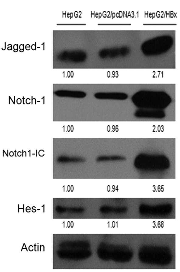
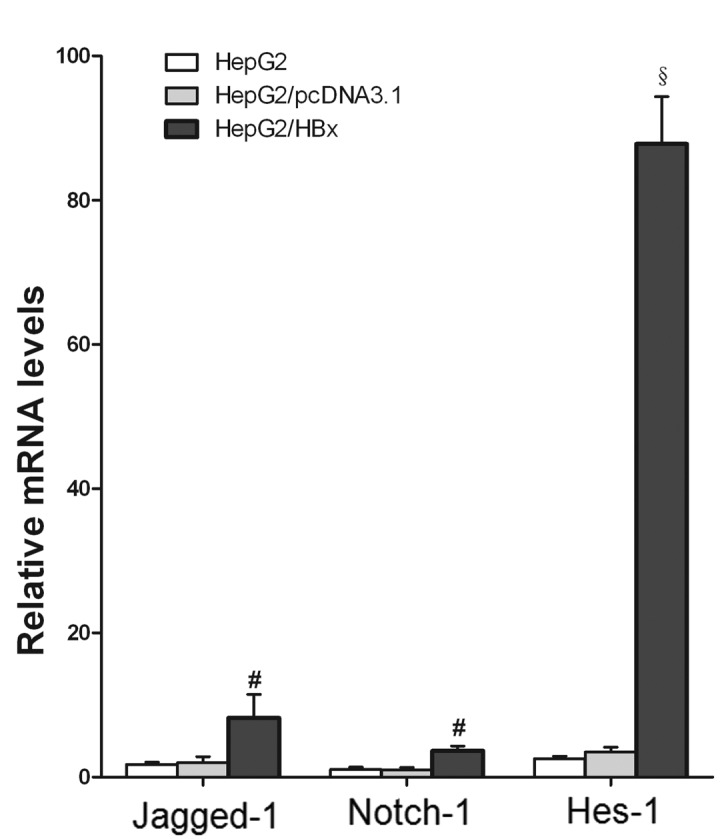
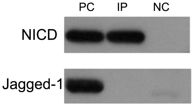
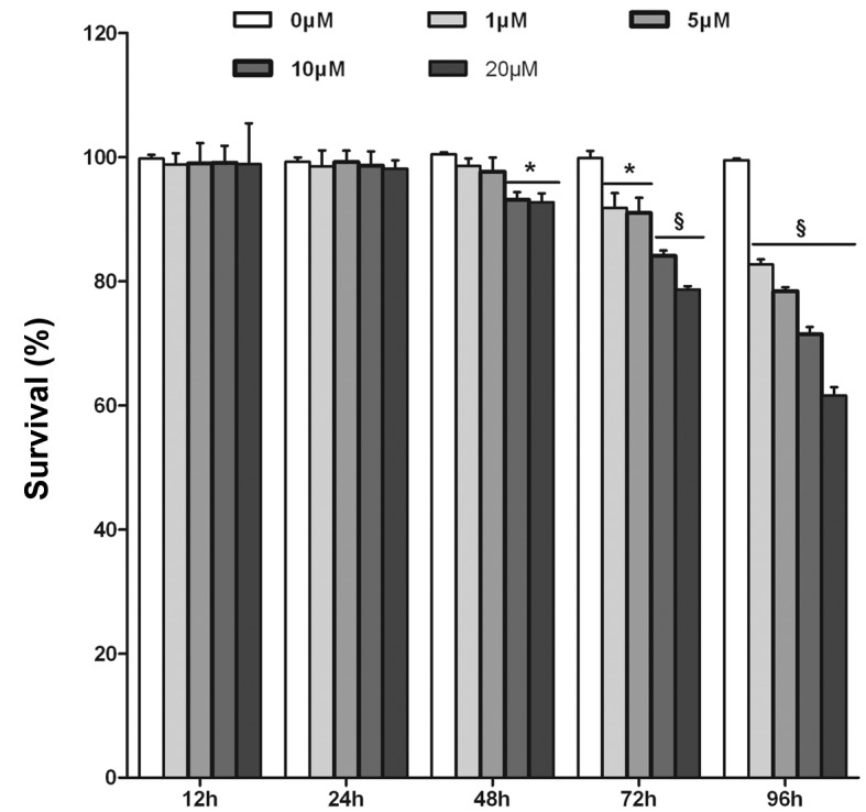
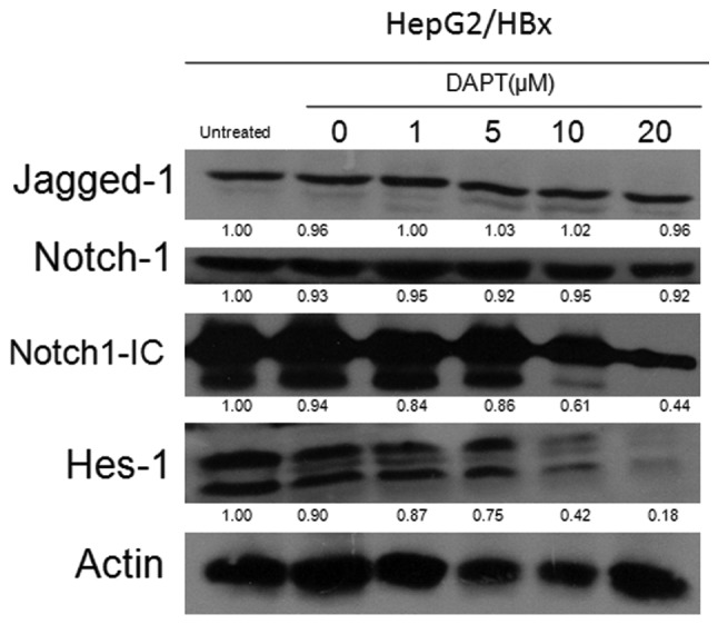
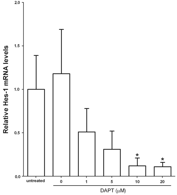
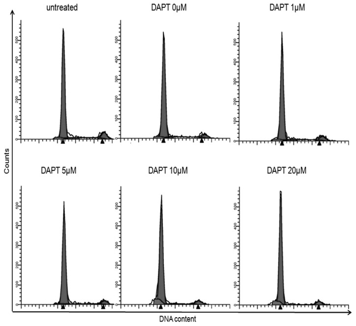
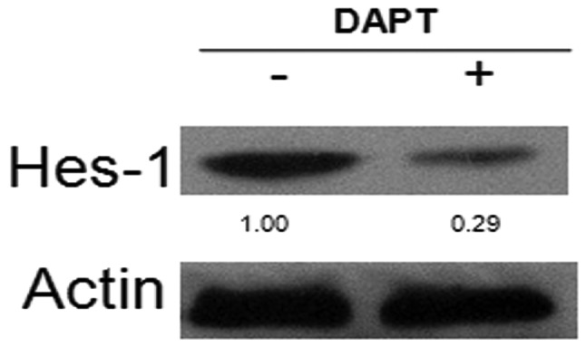
Hepatitis B virus X protein (HBx) plays a crucial role in the development of hepatocellular carcinoma (HCC), however, little is known about the mechanism. Here, we investigated the relationship between HBx and Notch signaling in HepG2 cells after they were transfected with the HBx gene. It was found that HBx upregulated the expression of Notch-1, Jagged-1 and Hes-1 at the transcriptional level by binding to the Notch-1 intracellular domain, which is congruent with the observations of enhanced malignant biological activities of HBx-transfected HepG2 cells compared with normal HepG2 cells. However, while Notch signaling was blocked, the HBx-induced abnormalities were partially reversed. These findings suggest that HBx may promote the progression of HCC via the activated Notch pathway.


Figures

Figure 1
Figure 1
Expression of Notch signaling pathway components and the physical interaction of NICD with HBx in HBx-expressing cells. (A) The protein levels of Notch signaling pathway components were detected using western blot analysis. Actin was used as an internal control for equal loading of samples. Representative blots shown were from three independent experiments with identical results. The relative ratios of each band were normalized to actin and are shown below each western blot band. (B) Jagged-1, Notch-1, Hes-1 mRNA expression levels were determined by quantitative real-time RT-PCR. Results are mean ± SEM of 3 independent experiments. #P<0.01 and §P<0.001 vs. control (HepG2 cells). (C) Confocal microscopy imaging of the co-localization of NICD with HBx in L02/HBx cells. The nuclei were stained with DAPI (blue) (i), HBx protein was stained with Cy3-conjugated goat anti-mouse IgG (red) (ii) and the protein of NICD was stained with FITC-conjugated goat anti-rabbit IgG (green) (iii). Yellow staining indicates overlapping areas of red and green fluorescent labels (iv). (D) Co-immunoprecipitation assay of the interaction of NICD and Jagged-1 with HBx in HepG2/HBx cells. The preparation of proteins was described in Materials and methods. All the proteins were analyzed by western blot analysis using anti-NICD, anti-Jagged-1. The data shown are representative of 3 independent experiments. PC, positive control; IP, co-immunoprecipitation of proteins treated with anti-HBx; NC, co-immunoprecipitation of proteins treated with non-immune mouse IgG (negative control).
Figure 1
Figure 1
Expression of Notch signaling pathway components and the physical interaction of NICD with HBx in HBx-expressing cells. (A) The protein levels of Notch signaling pathway components were detected using western blot analysis. Actin was used as an internal control for equal loading of samples. Representative blots shown were from three independent experiments with identical results. The relative ratios of each band were normalized to actin and are shown below each western blot band. (B) Jagged-1, Notch-1, Hes-1 mRNA expression levels were determined by quantitative real-time RT-PCR. Results are mean ± SEM of 3 independent experiments. #P<0.01 and §P<0.001 vs. control (HepG2 cells). (C) Confocal microscopy imaging of the co-localization of NICD with HBx in L02/HBx cells. The nuclei were stained with DAPI (blue) (i), HBx protein was stained with Cy3-conjugated goat anti-mouse IgG (red) (ii) and the protein of NICD was stained with FITC-conjugated goat anti-rabbit IgG (green) (iii). Yellow staining indicates overlapping areas of red and green fluorescent labels (iv). (D) Co-immunoprecipitation assay of the interaction of NICD and Jagged-1 with HBx in HepG2/HBx cells. The preparation of proteins was described in Materials and methods. All the proteins were analyzed by western blot analysis using anti-NICD, anti-Jagged-1. The data shown are representative of 3 independent experiments. PC, positive control; IP, co-immunoprecipitation of proteins treated with anti-HBx; NC, co-immunoprecipitation of proteins treated with non-immune mouse IgG (negative control).
Figure 2
Figure 2
(A) Effects of inhibition of Notch signaling on the growth of HepG2/HBx cells. The effect of DAPT on cell viability of HepG2/HBx cells was determined by the WST-8 assay as described in Materials and methods. Results are expressed as mean ± SEM of five samples. *P<0.05 vs. control (non-DAPT); §P<0.001 vs. control. (B and C) Notch signaling in HepG2/HBx cells by the γ-secretase inhibitor DAPT. (B) Inhibition of Notch signaling in HepG2/HBx cells with indicated concentrations of DAPT was assessed by immunoblotting. Cells were harvested after 48 h of treatment. Actin was used as an internal control for equal loading of samples. Representative blots shown are from three independent experiments with identical results. The relative ratios of each band were normalized to actin and are shown below each western blot band. (C) Hes-1 mRNA levels were assessed by qRT-PCR in DAPT-treated HepG2/HBx cells. Cells were harvested after 48 h of treatment. Data shown are mean ± SEM of representative experiment performed in triplicate. *P<0.01 vs. control (untreated cells).
Figure 2
Figure 2
(A) Effects of inhibition of Notch signaling on the growth of HepG2/HBx cells. The effect of DAPT on cell viability of HepG2/HBx cells was determined by the WST-8 assay as described in Materials and methods. Results are expressed as mean ± SEM of five samples. *P<0.05 vs. control (non-DAPT); §P<0.001 vs. control. (B and C) Notch signaling in HepG2/HBx cells by the γ-secretase inhibitor DAPT. (B) Inhibition of Notch signaling in HepG2/HBx cells with indicated concentrations of DAPT was assessed by immunoblotting. Cells were harvested after 48 h of treatment. Actin was used as an internal control for equal loading of samples. Representative blots shown are from three independent experiments with identical results. The relative ratios of each band were normalized to actin and are shown below each western blot band. (C) Hes-1 mRNA levels were assessed by qRT-PCR in DAPT-treated HepG2/HBx cells. Cells were harvested after 48 h of treatment. Data shown are mean ± SEM of representative experiment performed in triplicate. *P<0.01 vs. control (untreated cells).
Figure 3
Figure 3
Effects of the inhibition of Notch signaling in G0/G1 distribution, and apoptosis in HepG2/HBx cells. (A and B) The cell-cycle distribution of HepG2/HBx cells was examined by PI staining and flow cytometry synchronized by serum starvation, and treated with or without DAPT for 48 h. Results shown are from a representative experiment (A) or mean ± SEM of 3 experiments (B), each performed in triplicate. §P<0.05 vs. control (untreated cells). (C and D) Apoptosis of HepG2/HBx cells treated with or without DAPT for 48 h detected by Annexin-V and PI staining using FACS analysis. The percentage of cells that were Annexin-V positive, but PI negative were compared among different groups. Results shown are from a representative experiment (C) or mean ± SEM of 3 experiments (D), each performed in triplicate. #P<0.01 and §P<0.001 vs. control (untreated cells).
Figure 3
Figure 3
Effects of the inhibition of Notch signaling in G0/G1 distribution, and apoptosis in HepG2/HBx cells. (A and B) The cell-cycle distribution of HepG2/HBx cells was examined by PI staining and flow cytometry synchronized by serum starvation, and treated with or without DAPT for 48 h. Results shown are from a representative experiment (A) or mean ± SEM of 3 experiments (B), each performed in triplicate. §P<0.05 vs. control (untreated cells). (C and D) Apoptosis of HepG2/HBx cells treated with or without DAPT for 48 h detected by Annexin-V and PI staining using FACS analysis. The percentage of cells that were Annexin-V positive, but PI negative were compared among different groups. Results shown are from a representative experiment (C) or mean ± SEM of 3 experiments (D), each performed in triplicate. #P<0.01 and §P<0.001 vs. control (untreated cells).
Figure 4
Figure 4
Inhibition of Notch signaling in HepG2 cells. HepG2 cells were treated with DAPT at the final concentration of 10 μM or with 0.05% DMSO. (A) HepG2 cells (1×10⁴) were cultured in medium contained DAPT or DMSO for indicated times. Cell growth was assessed by the WST-8 assay as described in Materials and methods and plotted as a percentage of that of the control (untreated cells). Bars represent the means ± SEM of each time point from five samples. (B) HepG2 cells were harvested after 48 h of treatment with DAPT (10 μM) or vehicle control (0.05% DMSO). Inhibition of Notch signaling in HepG2 cells was monitored by immunoblotting for Hes-1 protein levels (+, DAPT; −, vehicle control).
Figure 4
Figure 4
Inhibition of Notch signaling in HepG2 cells. HepG2 cells were treated with DAPT at the final concentration of 10 μM or with 0.05% DMSO. (A) HepG2 cells (1×10⁴) were cultured in medium contained DAPT or DMSO for indicated times. Cell growth was assessed by the WST-8 assay as described in Materials and methods and plotted as a percentage of that of the control (untreated cells). Bars represent the means ± SEM of each time point from five samples. (B) HepG2 cells were harvested after 48 h of treatment with DAPT (10 μM) or vehicle control (0.05% DMSO). Inhibition of Notch signaling in HepG2 cells was monitored by immunoblotting for Hes-1 protein levels (+, DAPT; −, vehicle control).

Similar articles

Cited by

References

    1. El-Serag HB. Hepatocellular carcinoma: recent trends in the United States. Gastroenterology. 2004;127:S27–S34. - PubMed
    1. Llovet JM, Burroughs A, Bruix J. Hepatocellular carcinoma. Lancet. 2003;362:1907–1917. - PubMed
    1. Shibuya K, Mathers CD, Boschi-Pinto C, Lopez AD, Murray CJ. Global and regional estimates of cancer mortality and incidence by site: II. Results for the global burden of disease 2000. BMC Cancer. 2002;2:37. - PMC - PubMed
    1. Koike K. Hepatitis B virus HBx gene and hepatocarcinogenesis. Intervirology. 1995;38:134–142. - PubMed
    1. Lucito R, Schneider RJ. Hepatitis B virus X protein activates transcription factor NF-kappa B without a requirement for protein kinase C. J Virol. 1992;66:983–991. - PMC - PubMed

Publication types

MeSH terms

Substances