Editorial

Aβ Imaging: feasible, pertinent, and vital to progress in Alzheimer's disease

Victor L Villemagne et al. Eur J Nucl Med Mol Imaging. 2012 Feb.
No abstract available


Figures

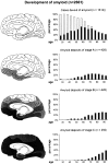
Fig. 1
Fig. 1
“Development of amyloid deposits in 2,661 nonselected autopsy cases. The first line displays the frequency of cases devoid of changes in relation to the total number of cases in the various age categories. The second, third, and fourth lines are similarly designed, and show the evolution of the AD-related changes. The dark areas of the columns refer to subgroups showing the presence of neurofibrillary changes.” (reproduced with permission from Neurobiol Aging, [22])
Fig. 2
Fig. 2
Correlations of the in vivo PiB distribution volume ratio (DVR) values with postmortem quantifications of [3H]PiB binding (E) and total insoluble Aβ (Aβ1-40 + Aβ1-42) peptide levels (F) in 19 brain regions from fresh-frozen tissue from the right hemisphere of the same Alzheimer’s disease subject. (reproduced with permission from Brain, [36])
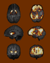
Fig. 3
Fig. 3
Comparison of the regional distribution of PiB retention in a presenilin-1 mutation carrier (left) and a sporadic case of AD (right). The MRI is shown in gray and PiB retention is overlaid in a hot metal scale on transaxial (top), sagittal (middle), and coronal images (bottom)

Comment on

Similar articles

Cited by

References

    1. Moghbel MC, Saboury B, Basu S, Metzler SD, Torigian DA, Långström B, Alavi A. Amyloid-beta imaging with PET in Alzheimer’s disease: is it feasible with current radiotracers and technologies? Eur J Nucl Med Mol Imaging 2011. doi:10.1007/s00259-011-1960-4. - PubMed
    1. Klunk WE, Wang Y, Huang GF, Debnath ML, Holt DP, Shao L, Hamilton RL, Ikonomovic MD, DeKosky ST, Mathis CA. The binding of 2-(4'-methylaminophenyl)benzothiazole to postmortem brain homogenates is dominated by the amyloid component. J Neurosci. 2003;23(6):2086–2092. - PMC - PubMed
    1. Fodero-Tavoletti MT, Rowe CC, McLean CA, Leone L, Li QX, Masters CL, Cappai R, Villemagne VL. Characterization of PiB binding to white matter in Alzheimer disease and other dementias. J Nucl Med. 2009;50(2):198–204. doi: 10.2967/jnumed.108.057984. - DOI - PubMed
    1. Klunk WE, Engler H, Nordberg A, Wang Y, Blomqvist G, Holt DP, Bergström M, Savitcheva I, Huang GF, Estrada S, Ausén B, Debnath ML, Barletta J, Price JC, Sandell J, Lopresti BJ, Wall A, Koivisto P, Antoni G, Mathis CA, Långström B. Imaging brain amyloid in Alzheimer’s disease with Pittsburgh Compound-B. Ann Neurol. 2004;55(3):306–319. doi: 10.1002/ana.20009. - DOI - PubMed
    1. Mathis CA, Wang Y, Holt DP, Huang GF, Debnath ML, Klunk WE. Synthesis and evaluation of 11C-labeled 6-substituted 2-arylbenzothiazoles as amyloid imaging agents. J Med Chem. 2003;46(13):2740–2754. doi: 10.1021/jm030026b. - DOI - PubMed

MeSH terms