. 2012 Mar;23(3):813-21.
doi: 10.1007/s10856-011-4545-7. Epub 2012 Jan 5.

Migration of intervertebral disc cells into dense collagen scaffolds intended for functional replacement

Affiliations

Migration of intervertebral disc cells into dense collagen scaffolds intended for functional replacement

Johannes L Bron et al. J Mater Sci Mater Med. 2012 Mar.

Abstract

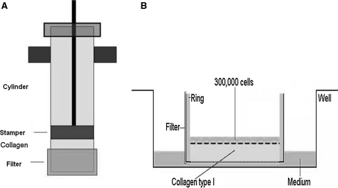
Invasion of cells from surrounding tissues is a crucial step for regeneration when using a-cellular scaffolds as a replacement of the nucleus pulposus (NP). The aim of current study was to assess whether NP and surrounding annulus fibrosus (AF) cells are capable of migrating into dense collagen scaffolds. We seeded freshly harvested caprine NP and AF cells onto scaffolds consisting of 1.5 and 3.0% type I collagen matrices, prepared by plastic compression, to assess cell invasion. The migration distance appeared both time and density dependent and was higher for NP (25%) compared to AF (10%) cells after 4 weeks. Migration distance was not enhanced by Hst-2, a peptide derived from saliva known to enhance fibroblast migration, and this was confirmed in a scratch assay. In conclusion, we revealed invasion of cells into dense collagen scaffolds and therewith encouraging first steps towards the use of a-cellular scaffolds for NP replacement.


Figures

Fig. 1
Fig. 1
Transmission electron microscopic pictures of the collagen scaffolds; a Uncompressed 0.5% (~5 mg/mL), b Compressed 1.5% (~15 mg/mL) and c Compressed 3% (~30 mg/mL). The spaces between individual collagen fibers can be estimated from the in-picture bar shown on the right below
Fig. 2
Fig. 2
Schematic presentation of the set up used for the migration experiments. First collagen scaffold are made by plastic compression in a cylinder with a cell culture insert used as a filter on the bottom (a). The filter is then transferred to a culture well and cells are seeded on top of the scaffolds (b)
Fig. 3
Fig. 3
Examples of 1.5% collagen scaffolds seeded with AF (a) and NP (b) cells after 28 days of migration (stain HE. Magnification ×20)
Fig. 4
Fig. 4
Graphic showing the results of the migration experiments of NP (white bars) and AF (black bars) cells after 14 and 28 days in 1.5 and 3% collagen matrices. After 14 days, no significant differences are observed. After 28 days, NP cells show a significantly higher migration compared to the AF cells. For both cell types the migration is higher in the 1.5 compared to the 3% collagen scaffolds
Fig. 5
Fig. 5
Surface area of 1, 5% collagen type I scaffolds after 1 week of incubation. (a AF cells, b NP cells. Stain DiI)
Fig. 6
Fig. 6
Graphic showing the results of the migration experiments of NP in a 1.5% collagen matrix with and without the addition of Hst-2 as a chemo attractant. The results with the negative enantiomer DHst-2 are also shown. The samples with Hst2 and Dhst-2 reveal a significantly decreased migration after 28 days
Fig. 7
Fig. 7
Images showing the results of Endo180 staining (HE counterstain) of 1.5% collagen scaffolds after 14 days of migration. a AF cells stained for Endo180 (brown color), b AF cells, negative control, c NP cells stained for Endo180 (brown color), d NP cells negative control
Fig. 8
Fig. 8
Graphic showing the results of the scratch assay. No significant differences are observed between the samples supplemented with Hst-2 compared to the negative control (D-Hst2) or serum free medium. The addition of 10% serum to the medium resulted in a significant increase in closure of the scratches
Fig. 9
Fig. 9
Images of cells during the scratch assay stained for F-actin (green) and the nuclei are stained with DAPI (blue). a NP cells outside the scratch, b NP cells inside the scratch, c AF cells outside the scratch, d AF cells inside the scratch
Fig. 10
Fig. 10
A confirmation scratch assay with human squamous carcinoma HO-1-N-1 cells reveals a significant enhanced cell migration after the addition of Hst-2

References

    1. Abbushi A, Endres M, Cabraja M, Kroppenstedt SN, Thomale UW, Sittinger M, Hegewald AA, Morawietz L, Lemke AJ, Bansemer VG, Kaps C, Woiciechowsky C. Regeneration of intervertebral disc tissue by resorbable cell-free polyglycolic acid-based implants in a rabbit model of disc degeneration. Spine. 2008;33:1527–1532. doi: 10.1097/BRS.0b013e3181788760. - DOI - PubMed
    1. Alini M, Li W, Markovic P, Aebi M, Spiro RC, Roughley PJ. The potential and limitations of a cell-seeded collagen/hyaluronan scaffold to engineer an intervertebral disc-like matrix. Spine. 2003;28:446–454. - PubMed
    1. Boyd LM, Carter AJ. Injectable biomaterials and vertebral endplate treatment for repair and regeneration of the intervertebral disc. Eur Spine J. 2006;15(Suppl 3):S414–S421. doi: 10.1007/s00586-006-0172-2. - DOI - PMC - PubMed
    1. Breuls RG, Jiya TU, Smit TH. Scaffold stiffness influences cell behavior: opportunities for skeletal tissue engineering. Open Orthop J. 2008;2:103–109. doi: 10.2174/1874325000802010103. - DOI - PMC - PubMed
    1. Bron JL, Helder MN, Meisel HJ, van Royen BJ, Smit TH. Repair, regenerative and supportive therapies of the annulus fibrosus: achievements and challenges. Eur Spine J. 2009;18:301–313. doi: 10.1007/s00586-008-0856-x. - DOI - PMC - PubMed

MeSH terms

LinkOut - more resources