. 2012 Mar 16;287(12):9360-75.
doi: 10.1074/jbc.M111.294546. Epub 2012 Jan 4.

Novel role for non-muscle myosin light chain kinase (MLCK) in hyperoxia-induced recruitment of cytoskeletal proteins, NADPH oxidase activation, and reactive oxygen species generation in lung endothelium

Affiliations

Novel role for non-muscle myosin light chain kinase (MLCK) in hyperoxia-induced recruitment of cytoskeletal proteins, NADPH oxidase activation, and reactive oxygen species generation in lung endothelium

Peter V Usatyuk et al. J Biol Chem. .

Abstract

We recently demonstrated that hyperoxia (HO) activates lung endothelial cell NADPH oxidase and generates reactive oxygen species (ROS)/superoxide via Src-dependent tyrosine phosphorylation of p47(phox) and cortactin. Here, we demonstrate that the non-muscle ~214-kDa myosin light chain (MLC) kinase (nmMLCK) modulates the interaction between cortactin and p47(phox) that plays a role in the assembly and activation of endothelial NADPH oxidase. Overexpression of FLAG-tagged wild type MLCK in human pulmonary artery endothelial cells enhanced interaction and co-localization between cortactin and p47(phox) at the cell periphery and ROS production, whereas abrogation of MLCK using specific siRNA significantly inhibited the above. Furthermore, HO stimulated phosphorylation of MLC and recruitment of phosphorylated and non-phosphorylated cortactin, MLC, Src, and p47(phox) to caveolin-enriched microdomains (CEM), whereas silencing nmMLCK with siRNA blocked recruitment of these components to CEM and ROS generation. Exposure of nmMLCK(-/-) null mice to HO (72 h) reduced ROS production, lung inflammation, and pulmonary leak compared with control mice. These results suggest a novel role for nmMLCK in hyperoxia-induced recruitment of cytoskeletal proteins and NADPH oxidase components to CEM, ROS production, and lung injury.


Figures
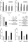
FIGURE 1.
FIGURE 1.
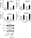
Role of nmMLCK in hyperoxia-mediated ROS and superoxide production in HPAECs. HPAECs grown on 35-mm dishes (∼90% confluence) were pretreated with increasing concentrations of ML-7 for 30 min and exposed to normoxia or hyperoxia for 3 h, and total ROS and superoxide accumulations were measured by DCFDA and hydroethidine immunofluorescence (A and B). In C, E, F, and G, HPAECs were transfected with scrambled (scRNA) or nmMLCK siRNA (50 nm) for 72 h as described under “Experimental Procedures.” Values for ROS and superoxide production are the mean ± S.D. from three independent experiments done in triplicate. *, significantly different from normoxia (p < 0.05); **, significantly different from untreated hyperoxia (p < 0.01). Total RNA was isolated from scrambled and nmMLCK siRNA-transfected cells, and real-time PCR was performed in a Light Cycler using SYBR Green QuantiTect (C). Data are from three independent experiments and are expressed as relative gene expression normalized to 18 S RNA. In D and E, cell lysates (20 μg of proteins) from ML-7, scrambled or nmMLCK siRNA-transfected cells were subjected to SDS-PAGE on 10% precast Tris-glycine gels and Western-blotted (IB) with anti-nmMLCK, phospho-MLC, and MLC antibodies. Shown is a representative blot of three independent experiments. In F and G, HPAECs were transfected with scrambled RNA or nmMLCK siRNA (50 nm) for 72 h, and cells were loaded with 10 μm DCFDA (F) or hydroethidine (G) for 30 min before exposure to normoxia or hyperoxia for 3 h. Formation of total ROS (F) and superoxide (G) was quantified as described above. Values are the mean ± S.D. of three independent experiments in triplicate. *, significantly different from normoxia (N) (p < 0.05); **, significantly different from hyperoxia exposure (HO) (p < 0.01).
FIGURE 2.
FIGURE 2.
Overexpression of nmMLCK potentiates hyperoxia-induced ROS and superoxide production in HPAECs. HPAECs grown on 35-mm dishes (∼60% confluence) were transfected with vector control or FLAG-tagged nmMLCK WT (1 μg/ml) using FuGENE HD (3 μg/ml) transfection reagent for 72 h as described under “Experimental Procedures.” A, cell lysates (20 μg of proteins) from vector control and FLAG-tagged nmMLCK transfected cells were subjected to SDS-PAGE and immunoblotted with anti-MLCK, anti-FLAG and anti-actin antibodies. A representative immunoblot from three independent experiments is shown. B and C, vector control and FLAG-tagged nmMLCK (WT)-transfected cells were loaded with 10 μm DCFDA (B) or hydroethidine (C) for 30 min before exposure to normoxia (N) or HO for 3 h. Formation of total ROS (B) and superoxide (C) were quantified as described under “Experimental Procedures.” Values are the mean ± S.D. of three independent experiments in triplicate. *, significantly different from normoxia (N) (p < 0.05); **, significantly different from vector control cells exposed to hyperoxia (HO) (p < 0.001).
FIGURE 3.
FIGURE 3.
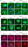
nmMLCK siRNA attenuates hyperoxia-induced phosphorylation of MLC and cortactin and actin rearrangement in HPAECs. HPAECs grown on 8-well slide chambers were transfected with scrambled RNA or nmMLCK siRNA (50 nm) for 72 h before exposure to either normoxia (N) or HO for 3 h, washed, fixed, permeabilized, and probed with anti-phospho-cortactin, anti-phospho-MLC antibodies, or phalloidin for actin staining. nmMLCK siRNA blunted hyperoxia-induced phosphorylation of cortactin and MLC near the cell periphery and enhanced actin stress fibers under both normoxia and hyperoxia as determined by immunofluorescent microscopy. A representative immunofluorescence image from three independent experiments is shown.
FIGURE 4.
FIGURE 4.
Down-regulation of nmMLCK with nmMLCK siRNA blunted hyperoxia-induced translocation and co-localization of p47phox with cortactin. HPAECs grown on 8-well slide chambers were transfected with scrambled siRNA or MLCK siRNA for 72 h then exposed to normoxia or hyperoxia (3 h), washed, fixed, permeabilized, probed with anti-cortactin or anti-p47phox antibodies, and examined by immunofluorescence microscopy using a 60× oil objective. The cortactin (red) and p47phox (green) images show matched cell fields for each condition. Exposure of cells to hyperoxia resulted in redistribution of cortactin and p47phox to the cell periphery, whereas MLCK siRNA blunted cortactin and p47phox redistribution and co-localization (yellow in merged images). A representative image from three independent experiments is shown.
FIGURE 5.
FIGURE 5.
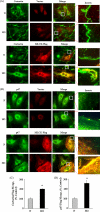
Overexpression of MLCK-FLAG wild type enhances hyperoxia-induced association of nmMLCK with cortactin and p47phox in cell periphery. HPAECs grown on 8-well slide chambers were transfected with vector control or FLAG-tagged MLCK wild type (1 μg/ml) and FuGENE HD (3 μg/ml) transfection reagent for 72 h and then exposed to either normoxia (N) or HO for 3 h. Cells were, washed, fixed, permeabilized, and probed with anti-cortactin (A), anti-p47phox (B), or anti-FLAG (A and B) antibodies and examined by immunofluorescence microscopy using a 60× oil objective. Exposure of cells to hyperoxia resulted in redistribution of cortactin and p47phox to the cell periphery with enhanced nmMLCK association and co-localization (yellow in merged images). Shown is a representative image from three independent experiments. C and D represent semiquantitation of the co-localization using an image analyzer showed an ∼2.0-fold increase in co-localization (yellow) between nmMLCK (red) and cortactin (green) and an ∼2.5-fold increase in co-localization between nmMLCK and p47phox.
FIGURE 6.
FIGURE 6.
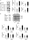
Down-regulation of nmMLCK with nmMLCK siRNA blunted the hyperoxia-induced association of cortactin with Src and p47phox and phosphorylation of cortactin, Src, and p47phox in HPAECs. A, HPAECs grown in 100-mm dishes to ∼90% confluence were exposed to either normoxia (N) or HO for 3 h. Cell lysates (500 μg proteins) from normoxia- or HO-exposed cells were immunoprecipitated (IP) with anti-nmMLCK antibody as described under “Experimental Procedures.” Immunoprecipitates were analyzed by 10% SDS-PAGE and probed with anti-nmMLCK, anti-cortactin, anti-Src, anti-p47phox, anti-actin, and anti-MLC antibodies. A representative blot from three independent experiments is shown. IB, immunoblot. B, HPAECs grown in 100-mm dishes to ∼50% confluence were transfected with scrambled (sc) RNA or nmMLCK siRNA (50 nm) for 72 h, and down-regulation of nmMLCK expression was verified by Western blotting as described under “Experimental Procedures.” Total cell lysates (500 μg of proteins) from scrambled and nmMLCK siRNA-treated cells were subjected to immunoprecipitation with anti-cortactin antibody, and immunoprecipitates were analyzed by 10% SDS-PAGE and probed with anti-cortactin (equal loading), anti-Src, anti-p47phox, anti-phosphotyrosine and anti-MLC antibodies. A representative blot from three independent experiments is shown.
FIGURE 7.
FIGURE 7.
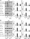
nmMLCK regulates hyperoxia-induced enrichment of cortactin, Src, and p47phox in caveolin-enriched microdomains. HPAECs grown in 100-mm dishes were transfected with scrambled (sc) RNA or nmMLCK siRNA (50 nm) for 72 h (A) or pretreated with ML-7 (1 μm, 30 min) (B) before exposure to either normoxia (N) or HO for 3 h, and caveolin-enriched microdomains were isolated as described under “Experimental Procedures.” Samples were then analyzed by 4–20% SDS-PAGE and probed with anti-cortactin, anti-Src, anti-p47phox, anti-MLC, anti-actin, anti-phospho-cortactin, anti-phospho-Src, and anti-phospho-MLC antibodies. Immunoblots (IB) were quantified by ImageJ software and expressed as a ratio to total caveolin-1. Values are the mean ± S.D. from three independent experiments. *, significantly different from normoxia (p < 0.05); **, significantly different from scrambled siRNA transfected cells exposed to hyperoxia.
FIGURE 8.
FIGURE 8.
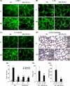
nmMLCK-KO mice exhibit reduced lung injury, leakage, and inflammation and ROS production in vivo. Male C57BL/6 WT or nmMLCK knock out (−/−) mice in the same background were exposed to either normoxia (N) or HO for 72 h. At the end of the experiment mice were anesthetized, BAL fluid was collected, lungs were perfused with fresh PBS several times, and whole lungs without trachea were paraffin-embedded. A–C, paraffin-embedded lung tissues from normoxia or HO animals were, sectioned, and immunostained with anti-phospho-MLC (A), anti-phospho-Src (B), or anti-phospho-cortactin (C) antibodies and examined under immunofluorescence microscopy using 60× oil objective. D, lung tissues from normoxia or HO-exposed animals were stored in formalin for 24 h before processing for staining with hematoxylin and eosin and lung morphology was evaluated using 40 X objective. E, BAL fluid collected from normoxia- or HO-exposed animals were subjected to cytospin, and differential cell counts were performed. Shown is a graphic representation of macrophages (M) and neutrophils (N) in BAL fluid from wild type and nmMLCK−/− mice exposed to normoxia or hyperoxia. Values are the mean ± S.D. from three independent experiments. *, significantly different from animals exposed to normoxia (p < 0.05); **, significantly different from animals exposed to normoxia (p < 0.01); ***, significantly different from wild type mice exposed to hyperoxia (p < 0.005). F and G, BAL fluid from mice exposed to normoxia or hyperoxia was analyzed for H2O2 (F) and total protein (G). Values are the mean ± S.D. from three independent experiments. *, significantly different from animals exposed to normoxia (p < 0.05); **, significantly different from wild type animals exposed to hyperoxia (p < 0.01).
FIGURE 9.
FIGURE 9.
Overexpression of FLAG-tagged nmMLCK wild type in nmMLCK-deficient mouse lung ECs restores hyperoxia-induced ROS/O2˙̄ production. Mouse lung endothelial cells isolated from 4-5-week-old C57BL/6 and nmMLCK−/− null mice were isolated using collagenase A as described under “Experimental Procedures.” In A and B, lungs ECs grown to ∼80% confluence from wild type and nmMLCK−/− mice were exposed to normoxia or hyperoxia for 3 h, and total ROS and superoxide generated in cells were quantified as described under “Experimental Procedures.” Values for ROS and superoxide production are the mean ± S.D. from three independent experiments done in triplicate. *, significantly different from normoxia (p < 0.01); **, significantly different from untreated hyperoxia (p < 0.05). In C and D, lung ECs from nmMLCK−/− null mice were infected with vector control or adenoviral human nmMLCK (AdMLCK (WT)) (5 pfu/cell) in EBM-2 complete medium for 24 h before exposure to normoxia or hyperoxia for 3 h. Total ROS and superoxide accumulation in cells were measured by DCFDA and hydroethidine (HE) fluorescence. Values for ROS and superoxide production are the mean ± S.D. from three independent experiments done in triplicate. *, significantly different adenoviral nmMLCK wild type-infected cells exposed to normoxia (p < 0.01). In E, cell lysates (20 μg proteins) from wild type, nmMLCK−/−, and FLAG-tagged human nmMLCK-infected mouse lung ECs were subjected to SDS-PAGE and immunoblotted with anti-MLCK, anti-FLAG, and anti-actin antibodies. A representative immunoblot (IB) from three independent experiments is shown. Immunoblots were analyzed by densitometry and quantified.
FIGURE 10.
FIGURE 10.
Overexpression of FLAG-tagged nmMLCK wild type in nmMLCK-deficient mouse lung ECs restores hyperoxia-induced redistribution of p-MLC, p-Src, and p-cortactin to cell periphery. Mouse lung ECs isolated from 4–5-week-old C57BL/6 wild type and nmMLCK−/− mice were grown on 8-well slide chambers to ∼80% confluence. In some experiments ECs isolated from nmMLCK−/− null mice were infected with vector control or adenoviral human nmMLCK wild type (AdMLCK, 5 pfu/cell) for 24 h. Cells were exposed to normoxia (N) or HO for 3 h, washed, fixed, permeabilized, and probed with anti-phospho-MLC (A), anti-phospho-Src (B), or anti-phospho-cortactin (C) antibodies and examined by immunofluorescence microscopy using a 60× oil objective. A representative image from three independent experiments is shown.
FIGURE 11.
FIGURE 11.
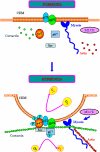
Proposed model on involvement of ∼214-kDa nmMLCK in assembly and activation of non-phagocytic NADPH oxidase and ROS/superoxide production in lung endothelium. As depicted in the model, both cortactin and p47phox are diffused throughout the cell and mostly co-localized to the same intracellular compartment. Upon exposure to hyperoxia, nmMLCK facilitates the assembly and association of cortactin, p47phox, Src, and other components to the caveolin-enriched microdomains at the cell periphery for enhanced ROS/superoxide production.

Similar articles

Cited by

References

    1. Frey R. S., Ushio-Fukai M., Malik A. B. (2009) NADPH oxidase-dependent signaling in endothelial cells. Role in physiology and pathophysiology. Antioxid. Redox Signal. 11, 791–810 - PMC - PubMed
    1. Pendyala S., Usatyuk P. V., Gorshkova I. A., Garcia J. G., Natarajan V. (2009) Regulation of NADPH oxidase in vascular endothelium. The role of phospholipases, protein kinases, and cytoskeletal proteins. Antioxid. Redox Signal. 11, 841–860 - PMC - PubMed
    1. Griffith B., Pendyala S., Hecker L., Lee P. J., Natarajan V., Thannickal V. J. (2009) NOX enzymes and pulmonary disease. Antioxid. Redox Signal. 11, 2505–2516 - PMC - PubMed
    1. Li J. M., Shah A. M. (2002) Intracellular localization and preassembly of the NADPH oxidase complex in cultured endothelial cells. J. Biol. Chem. 277, 19952–19960 - PubMed
    1. Touyz R. M., Yao G., Quinn M. T., Pagano P. J., Schiffrin E. L. (2005) p47phox associates with the cytoskeleton through cortactin in human vascular smooth muscle cells. Role in NAD(P)H oxidase regulation by angiotensin II. Arterioscler. Thromb. Vasc. Biol. 25, 512–518 - PubMed

Publication types

MeSH terms

LinkOut - more resources