. 2012 Feb 24;287(9):6406-20.
doi: 10.1074/jbc.M111.301622. Epub 2012 Jan 4.

Molecular determinants of enterovirus 71 viral entry: cleft around GLN-172 on VP1 protein interacts with variable region on scavenge receptor B 2

Affiliations

Molecular determinants of enterovirus 71 viral entry: cleft around GLN-172 on VP1 protein interacts with variable region on scavenge receptor B 2

Pan Chen et al. J Biol Chem. .

Abstract

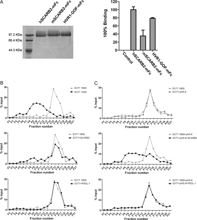
Enterovirus 71 (EV71) is one of the major pathogens that cause hand, foot, and mouth disease outbreaks in young children in the Asia-Pacific region in recent years. Human scavenger receptor class B 2 (SCARB2) is the main cellular receptor for EV71 on target cells. The requirements of the EV71-SCARB2 interaction have not been fully characterized, and it has not been determined whether SCARB2 serves as an uncoating receptor for EV71. Here we compared the efficiency of the receptor from different species including human, horseshoe bat, mouse, and hamster and demonstrated that the residues between 144 and 151 are critical for SCARB2 binding to viral capsid protein VP1 of EV71 and seven residues from the human receptor could convert murine SCARB2, an otherwise inefficient receptor, to an efficient receptor for EV71 viral infection. We also identified that EV71 binds to SCARB2 via a canyon of VP1 around residue Gln-172. Soluble SCARB2 could convert the EV71 virions from 160 S to 135 S particles, indicating that SCARB2 is an uncoating receptor of the virus. The uncoating efficiency of SCARB2 significantly increased in an acidic environment (pH 5.6). These studies elucidated the viral capsid and receptor determinants of enterovirus 71 infection and revealed a possible target for antiviral interventions.


Figures

FIGURE 1.
FIGURE 1.
Single round EV71 infection system. A, schematic map of the EV71 viral RNA genome, replication-competent EV71(FY)-EGFP viral genome, and EV71 replicon and capsid expresser for the single round EV71 infection system, EV71(FY)-Luc. EV71(FY)-EGFP is a replication-competent virus with the EGFP reporter gene inserted between the 5′-UTR and VP4. An EV71 2A protease recognition site (-AITTL-) was inserted between EGFP and VP4. For the single round EV71 infection system (EV71(FY)-Luc), the EV71 capsid expresser was used to express all the structural capsid genes in trans, an EGFP gene was inserted upstream of the EV71(FY) capsid gene, and all other EV71 viral genes were deleted. The EGFP was separated from the structural genes by a 2A self-cleavage site. EV71 replicon was produced by replacing the capsid coding region with a firefly luciferase reporter gene in the full-length EV71(FY) genome, and a T7 promoter was placed at the 5′-end for transcription in vitro. B, characterization of EV71 pseudotype virus by ultracentrifugation. Both wild type EV71(FY) and pseudotype virus EV71(FY)-Luc were applied to sucrose gradient ultracentrifugation, and the copy number of viral genomic RNA in each fraction was subsequently determined by quantitative RT-PCR. EV71(FY) virus was quantified with primers located in VP1. Specific primers for firefly luciferase reporter gene were used for quantifying pseudotype EV71(FY)-Luc virus. The data shown are representative of three independent experiments. Error bars represent S.D. between duplicate samples analyzed in the experiment. C, inhibition of EV71(FY)-Luc pseudotype viral infection of RD cells. EV71 pseudotype virus was incubated with patient anti-EV71 serum samples at 4 °C for 1 h prior to infection of RD cells in a 96-well plate in a total volume of 100 μl in quadruplicate. Luciferase activity was measured 24 h postinfection. Non-linear regression analysis of the neutralization curve of anti-EV71 serum was conducted using GraphPad Prism. N3, N12, and N25 are convalescent sera from different patients that showed low, medium, and high neutralization activity, respectively, against EV71 infection in a classic plaque reduction assay. IRES, internal ribosome entry site.
FIGURE 2.
FIGURE 2.
SCARB2 from diverse species supports EV71 infection with various efficiencies. A, SCARB2 knockdown RD (RD/SCARB2 kd) cell line. Left panel, the SCARB2 mRNA levels in RD cells, stable RD/SCARB2 kd cells, and control RD cells treated with an unrelated shRNA were determined by real time PCR. The SCARB2 mRNA levels for each cell line were normalized using GAPDH as an internal control. Right panel, relative binding efficiency of EV71 pseudotype virus EV71(FY)-Luc to RD and RD/SCARB2 kd cells. The data are shown as averages and S.D. from triplicates analyzed in one experiment, which is representative of three independent experiments. B, SCARB2s from human and horseshoe bat supported more efficient EV71(FY)-EGFP infection than those from mouse and hamster. Human SCARB2 (SCARB2smut that carries nonsense nucleotide mutations to mismatch shRNA target sequence) and SCARB2s of horseshoe bat, mouse, and hamster, respectively, were transiently transfected into a stable SCARB2 kd cell line. 24 h post-transfection, cells were infected with EV71(FY)-EGFP viruses, and infected cells were photographed 24 h postinfection. C, single round EV71(FY)-Luc infection of cells complemented with SCARB2 from human, mouse, hamster, and horseshoe bat. BHK21 cells were transfected with plasmids encoding SCARB2 (fused with a C9 tag at the C terminus) from different species as indicated, and the cells were then infected with EV71(FY)-Luc reporter virus. Luciferase activity was determined 24 h postinfection, and the percentage of infectivity was calculated and normalized to the infectivity of human SCARB2-transfected cells considered as 100%. Infection of EV71(FY)-Luc in untransfected BHK21 cells was used as a control. The data reflect the means with S.D. bars from quadruplicate data points within one experiment. Similar results were obtained from three independent experiments. D, cell surface expression of SCARB2s. BHK21 cells from the same transfections as in C were harvested 24 h post-transfection, cell surface molecules were biotinylated, and cells were lysed. The biotinylated SCARB2s were then pulled down with a C9-specific antibody, 1D4, from the cell lysate and subsequently detected with HRP-streptavidin by Western blot. Untransfected BHK21 cells were used as a control.
FIGURE 3.
FIGURE 3.
Identification of critical residues on SCARB2 for EV71 infection. A, schematic diagram of chimeric SCARB2 proteins. Swap mutants of human SCARB2 with residues from mouse SCARB2 at the highly variable regions (HVR1, HVR2, HVR3, and HVR4, respectively) were generated by replacing the human SCARB2 sequence (black) with the murine counterpart (white). The positions of the amino acids exchanged are indicated. Mouse SCARB2 with three (WHF), four (IESV), or seven (HVR1) GOF mutant residues was constructed by replacing the corresponding murine residues with their human counterparts on a mouse SCARB2 expression plasmid. All SCARB2s contain a C9 tag at the C terminus. TM, transmembrane domain. B, amino acid alignment of the HVR1 region of SCARB2s from four different species and GOF mutants of mouse SCARB2. GOF mutants are the three- (WHF), four- (IESV) or seven-residue (HVR1) mutations in the HVR1 region. Residues changed from mouse to human are indicated with underlines. C and D, swapping the HVR1 of human SCARB2 with the mouse counterpart interferes with EV71 infection. RD SCARB2/kd cells transfected with plasmids encoding human SCARB2, mouse SCARB2, or human SCARB2 HVR1–4 LOF mutants were infected with replication-competent EV71(FY)-GFP. Infected cells were photographed 18 h postinfection (C). Infections of EV71(FY)-Luc pseudotype virus were determined on BHK21 cells transfected with various SCARB2s. Firefly luciferase reporter activity was measured 24 h postinfection (D, lower panel), and the surface expression of these SCARB2 variants was also examined (D, upper panel). All SCARB2s contained a C9 tag at the C terminus. Untransfected BHK21 cells were used as a control in D. E and F, introduction of seven residues of human SCARB2 into mouse SCARB2 converts mouse SCARB2 to an efficient EV71 receptor for viral infection. EV71(FY)-GFP infection (E) was recorded as in C. For EV71(FY)-Luc infection (F), surface expression of SCARB2 mutants was determined for BHK21 cells transfected with SCARB2s (F, upper panel). EV71(FY)-Luc infection in these cells was measured 24 h postinfection (F, lower panel). Untransfected BHK21 cells were used as a control in F, and the infectivity of EV71(FY)-Luc in BHK21 cells transfected with hSCARB2 was set as 100%. All the data shown represent three or more experiments. Cell surface expression of SCARB2s was determined similarly to that in Fig. 2D. For both D and F, the data are representative of three independent experiments. Error bars represent S.D. from quadruplicate samples analyzed within the experiment.
FIGURE 4.
FIGURE 4.
EV71 enters cells via canyon around Gln-172 residue on VP1. A, effect of mutations on surface-exposed VP1 residues for EV71 infection of RD cells. B, effect of VP1 mutants on viral binding activity in RD cells. C, mutagenesis analysis of VP1 around Gln-172 revealed a cluster of residues involved in the interactions between VP1 and SCARB2 as shown by reduction in viral binding and RD cell infectivity. Upper panel, RNA genome copies of each mutant virion were examined with qRT-PCR. Middle panel, mutant virus binding to RD cells. The RD cell-bound EV71(FY)-Luc virions were quantified by RT-qPCR, and binding of mutant virus was normalized to the pseudotype virus bearing wild type VP1. Bottom panel, mutant viral infectivity measurement. EV71(FY)-Luc pseudotype mutant viruses were used to infect RD cells in quadruplicate, and viral infectivity was measured 24 h postinfection by luciferase reporter. Infectivity of mutant virus was normalized to the wild type EV71(FY)-Luc viruses. D, Western blot analysis of VP1 proteins on mutant EV71 pseudovirions. The same amounts of pseudovirions (normalized by viral RNA genome copies quantified by qRT-PCR) bearing mutant VP1 as indicated were examined for VP1 protein levels on the virions by Western blot. Pseudovirions were concentrated by ultracentrifugation, and VP1 proteins were detected by mouse anti-EV71 polyclonal serum. E, SCARB2-mFc pulldown assay on EV71 pseudovirions carrying mutant VP1. 1 μg of purified SCARB2-mFc fusion protein was incubated with ∼5 × 10⁸ pseudovirions bearing wild type or mutant VP1 at 4 °C for 4 h in the presence of protein A beads. After extensive washing with PBS three times, the bound virions were quantified by RT-qPCR. The data shown in A–E are representative of 2–3 independent experiments for each panel. Error bars represent S.D. from triplicate samples analyzed within one experiment. F, the potential binding interfaces between the canyon on EV71 VP1 and SCARB2 were mapped on the three-dimensional architecture of EV71 virus capsid. The capsid architecture shown was built by replacing the VP1 on the cryo-EM structure of poliovirus (Protein Data Bank code 2PLV) with our modeled VP1 structure. The EV71 VP1 residues whose alteration substantially decreased SCARB2 binding are colored in red (Gln-152, Arg-166, Trp-171, Thr-173, Gln-172, Thr-175, Asn-176, Ser-178, Phe-180, and Arg-236, respectively).
FIGURE 5.
FIGURE 5.
SCARB2 is an uncoating receptor of EV71. A, pulldown assay of EV71 with soluble SCARB2-mFc variants. Left panel, SDS-PAGE of purified SCARB2-mFc fusion proteins with the extracellular region of human or mouse SCARB2 or HVR1-GOF-mFc. Right panel, 1 μg of SCARB2-mFc proteins shown on the left was incubated with 1 μl of purified EV71(FY) wild type virus (∼5 × 10⁸ virions) at 4 °C for 4 h in the presence of protein A beads. After extensive washing with PBS three times, the bound virions were quantified by RT-qPCR. 1 μg of unrelated mFc fusion protein (coronavirus HKU1 S1-mFc) was used as a control. All data points show means and S.D. (error bars) for duplicate samples from a representative experiment (n = 3). B and C, virion flotation assay with sucrose density gradient ultracentrifugation. Purified virus (∼5 × 10⁹) was incubated with 5 μg of hSCARB2-mFc or hPSGL1-mFc followed by incubation at 37 °C for 100 min at neutral pH (B) or pH 5.6 (C) and then loaded onto a 15–30% discontinuous sucrose gradient for ultracentrifugation at 40,000 rpm for 70 min at 10 °C in a Beckman MLS-50 rotor. 20 μl of each fraction of a total 21 fractions was subjected to RT-qPCR to quantify virus in each fraction. The 135 S reference particle was made in vitro from native 160 S virions by heating for 3 min in a low salt buffer (4 mm CaCl2, 20 mm HEPES, pH 7.4).

References

    1. Racaniello V. R. (2007) in Fields Virology (Knipe D. M., Howley P. M., Griffin D. E., Lamb R. A., Martin M. A., Roizman B., Straus S. E., eds) 5 Ed., pp. 795–838, Vol. 1, Lippincott Williams & Wilkins, Philadelphia
    1. Huang C. C., Liu C. C., Chang Y. C., Chen C. Y., Wang S. T., Yeh T. F. (1999) Neurologic complications in children with enterovirus 71 infection. N. Engl. J. Med. 341, 936–942 - PubMed
    1. Vallet S., Legrand Quillien M. C., Dailland T., Podeur G., Gouriou S., Schuffenecker I., Payan C., Marcorelles P. (2009) Fatal case of enterovirus 71 infection, France, 2007. Emerg. Infect. Dis. 15, 1837–1840 - PMC - PubMed
    1. Lu M., Meng G., He Y. X., Zheng J., Liao S. L., Zhong Y. F., Zhao X. S., Shao H. Q., Wang Y. P., Gao Z. C., Gao Z. F. (2009) Pathology of enterovirus 71 infection: an autopsy study of 5 cases. Zhonghua Bing Li Xue Za Zhi 38, 81–85 - PubMed
    1. Lum L. C., Wong K. T., Lam S. K., Chua K. B., Goh A. Y. (1998) Neurogenic pulmonary oedema and enterovirus 71 encephalomyelitis. Lancet 352, 1391. - PubMed

Publication types

MeSH terms