. 2012 Apr 12;86(4):110.
doi: 10.1095/biolreprod.111.097501. Print 2012 Apr.

The autoimmune regulator prevents premature reproductive senescence in female mice

Affiliations

The autoimmune regulator prevents premature reproductive senescence in female mice

Susmita Jasti et al. Biol Reprod. .

Abstract

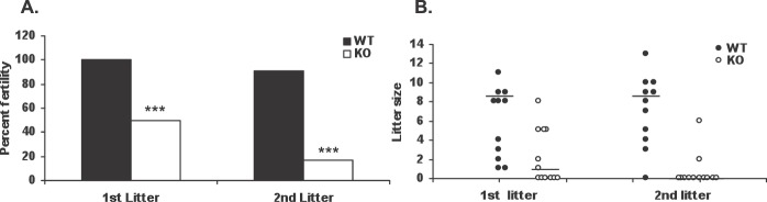
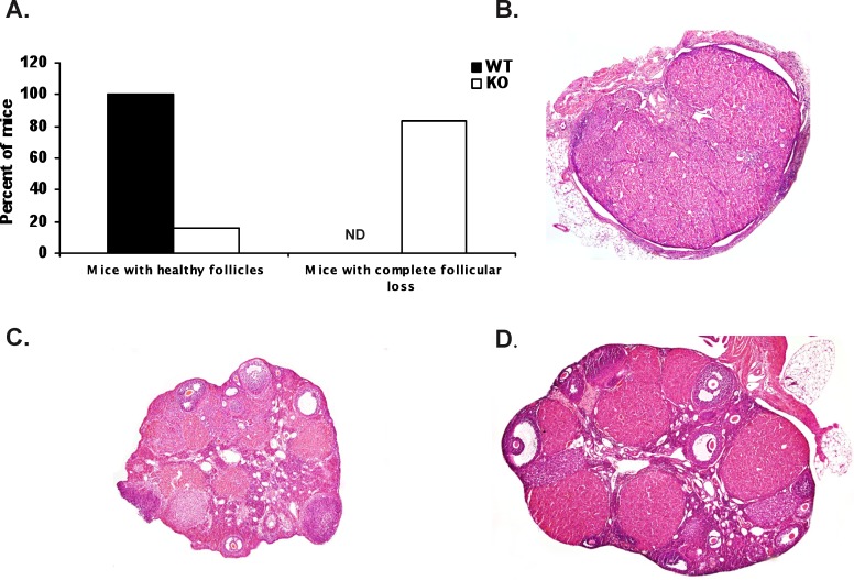
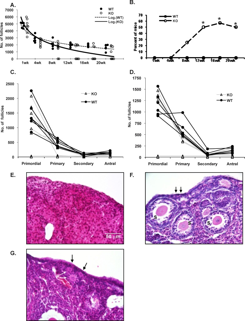
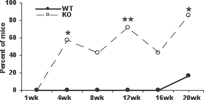
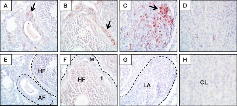
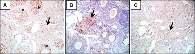
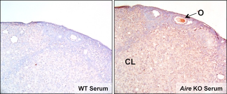
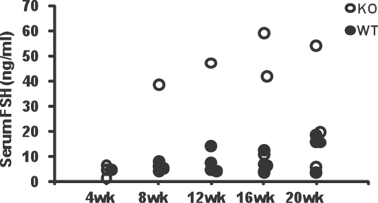
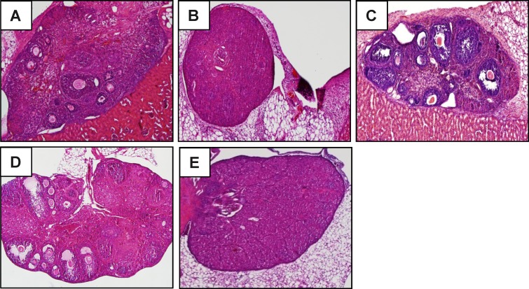
Loss-of-function mutations in the autoimmune regulator (AIRE) gene are responsible for autoimmune polyglandular syndrome type 1 (APS-1), which commonly manifests as infertility in women. AIRE is a transcriptional regulator that promotes expression of tissue-restricted antigens in the thymus, including antigens specific to the ovary. Thymic expression of ovarian genes under AIRE's control may be critical for preventing ovarian autoimmune disease. Because mice lacking Aire are an important APS-1 model, we examined the reproductive properties of female Aire-deficient (Aire(-/-)) mice. Female Aire(-/-) mice on the BALB/c background were examined for reproductive parameters, including fertility, litter sizes, and ovarian follicular reserves. Although delayed puberty was observed in Aire(-/-) mice, all mice entered puberty and exhibited mating behavior. Only 50% of Aire(-/-) females gave an initial litter, and only 16% were able to produce two litters. Ovarian histopathologic examination revealed that 83% of previously bred females lost all ovarian follicular reserves. Among virgin females, follicular depletion was observed in 25% by 8 wk, and by 20 wk, 50%-60% of mice lost all follicles. This was associated with elevated serum follicle-stimulating hormone level and ovarian infiltration of proliferating CD3+ T lymphocytes. Ovulation rates of 6-wk-old Aire(-/-) mice were reduced by 22%, but this difference was not statistically significant. Finally, transplantation experiments revealed that follicular loss depended on factors extrinsic to the ovary. These results suggest that immune-mediated ovarian follicular depletion is a mechanism of infertility in Aire(-/-) mice. The results have important implications in the pathogenesis of ovarian autoimmune disease in women.


Figures

FIG. 1
FIG. 1
Influence of Aire deficiency on female fertility. Wild-type (WT) and Aire-deficient (KO) mice were monitored across two consecutive litters as described in Materials and Methods. A) Percentage of WT control and KO mice giving rise to viable litters. B) Litter size. Each circle represents the litter size of an individual dam. Bars represent the median. ***P < 0.001 by chi-square analysis.
FIG. 2
FIG. 2
Ovarian phenotype in parous Aire-deficient (KO) mice. Mice were followed for two consecutive litters, and at euthanization, ovaries were harvested and processed for histological analysis. A) Percentage of KO and wild-type (WT) mice with intact and depleted follicular reserves. BD) Histological sections of ovaries from representative KO (B and C) and WT (D) mice exhibiting follicular loss (B) and intact follicles (C and D). ND, nondetected. Original magnification ×100.
FIG. 3
FIG. 3
Impact of Aire deficiency on ovarian follicular reserves. A) Ovarian follicular reserves were counted from virgin Aire-deficient (KO) and wild-type (WT) mice as described in Materials and Methods. Data are expressed as numbers of healthy follicles per ovary. Each circle represents the number of follicles from an individual mouse. Dashed and solid lines represent trend lines for the WT control and KO groups, respectively. The x-axis represents age of mice in weeks. B) Percentage of WT and KO mice exhibiting complete follicular loss as a function of age. C and D) Numbers of follicles of different stages found in WT and KO mice at 8 wk (C) and 12 wk (D) of age. Each line represents an individual mouse. EG) Representative images of ovaries from 8-wk-old, nulliparous KO (E and F) and WT (G) mice. Arrows indicate primordial follicles. P, primary follicles; S, secondary follicles. *P < 0.05. Bar = 50 μm.
FIG. 4
FIG. 4
Proportion of mice exhibiting CD3+ T cell infiltration into ovaries of Aire-deficient (KO) and wild-type (WT) mice. *P < 0.05, **P < 0.01 as compared to WT controls.
FIG. 5
FIG. 5
Infiltration of CD3+ T cells into ovaries of Aire-deficient mice. Ovaries of wild-type (not shown) and Aire-deficient (AH) mice were stained by immunohistochemistry using an anti-mouse CD3 antibody. Reddish-brown staining represents positive staining (arrows); sections were counterstained with hematoxylin (blue). T cells are seen surrounding atretic follicles (AF; A), surrounding healthy follicles (HF; B), organized into lymphoid aggregates (LA; C), and infiltrating corpora lutea (CL; D). Corresponding isotype controls (EH) are also shown. te, theca externa; ti, theca interna. Original magnification ×600.
FIG. 6
FIG. 6
CD3+ cells proliferate in situ within the ovary. Serial sections of ovaries of Aire-deficient (AC) and wild-type (not shown) mice were stained with MKI67 (A), CD3 (B), and isotype control (C) antibodies. Arrows show areas of intense staining by both antibodies. F, follicles. Original magnification ×200 (A and B) and ×400 (C).
FIG. 7
FIG. 7
Ovary-reactive autoantibodies are produced in Aire-deficient mice. Ovaries from Rag1-deficient mice were stained by immunohistochemistry using serum from wild-type or Aire-deficient mice. CL, corpus luteum; O, oocyte. Original magnification ×20.
FIG. 8
FIG. 8
Serum FSH concentrations (ng/ml) in Aire-deficient (KO) and wild-type (WT) mice. Each circle represents FSH concentration in an individual mouse. The x-axis represents age in weeks.
FIG. 9
FIG. 9
Follicular loss is independent of AIRE expression intrinsic to the ovary. Aire-deficient (A and B) and wild-type (C) mice were engrafted with a donor wild-type ovary under the kidney capsule and euthanized after 10 days. Grafts and endogenous ovaries were harvested for histological analysis. AC represent the grafted ovary; D and E represent the endogenous ovary of the Aire-deficient recipients. Original magnification ×100.

Comment in

Similar articles

Cited by

References

    1. Forges T, Monnier-Barbarino P, Faure GC, Bene MC. Autoimmunity and antigenic targets in ovarian pathology. Hum Reprod Update 2004; 10: 163 175 - PubMed
    1. Conway GS, Kaltsas G, Patel A, Davies MC, Jacobs HS. Characterization of idiopathic premature ovarian failure. Fertil Steril 1996; 65: 337 341 - PubMed
    1. Kyewski B, Klein L. A central role for central tolerance. Annu Rev Immunol 2006; 24: 571 606 - PubMed
    1. Starr TK, Jameson SC, Hogquist KA. Positive and negative selection of T cells. Annu Rev Immunol 2003; 21: 139 176 - PubMed
    1. Smith KM, Olson DC, Hirose R, Hanahan D. Pancreatic gene expression in rare cells of thymic medulla: evidence for functional contribution to T cell tolerance. Int Immunol 1997; 9: 1355 1365 - PubMed

Publication types

MeSH terms