. 2012 Jan 4;32(1):183-93.
doi: 10.1523/JNEUROSCI.1305-11.2012.

Transgenic mouse model expressing the caspase 6 fragment of mutant huntingtin

Affiliations

Transgenic mouse model expressing the caspase 6 fragment of mutant huntingtin

Elaine Waldron-Roby et al. J Neurosci. .

Abstract

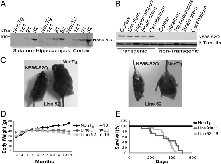
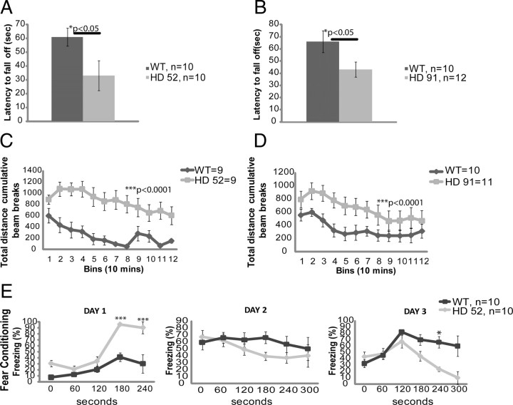
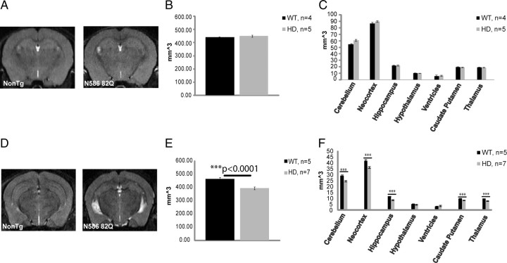
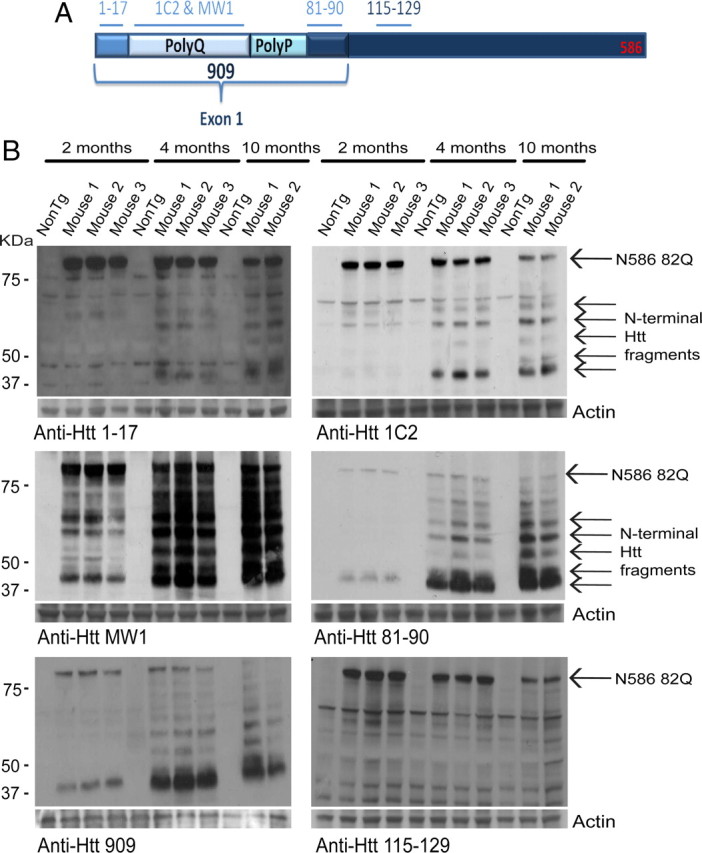
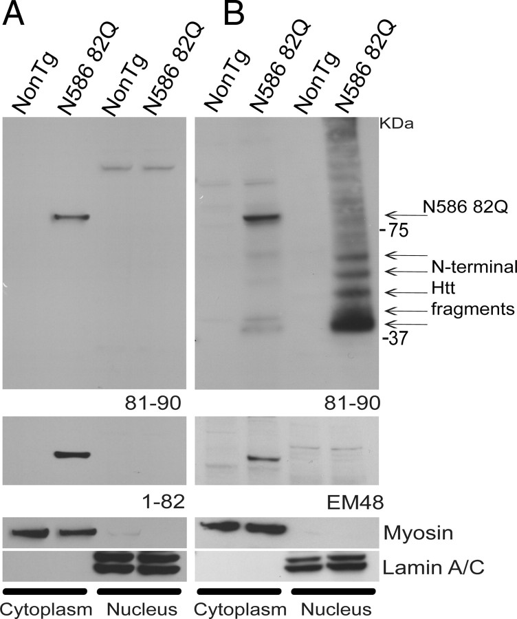
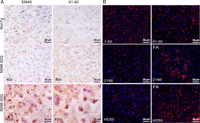
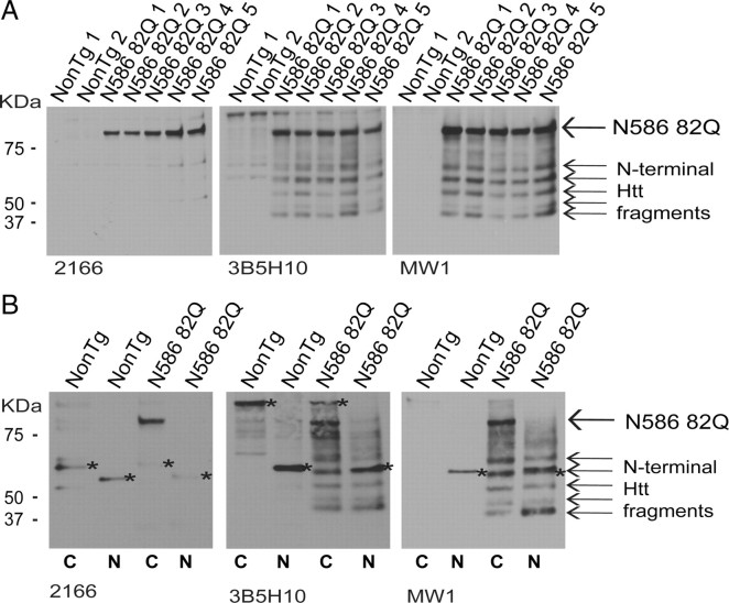
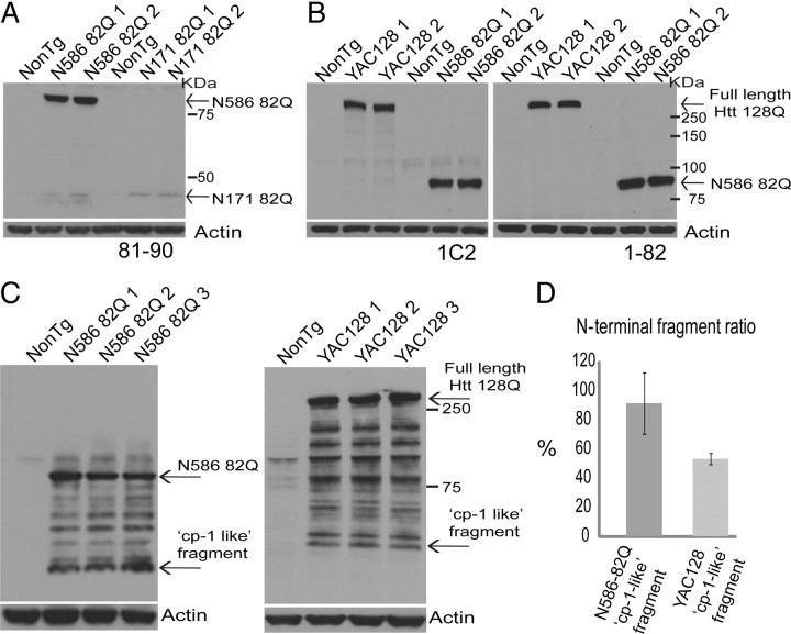
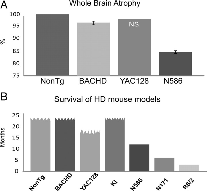
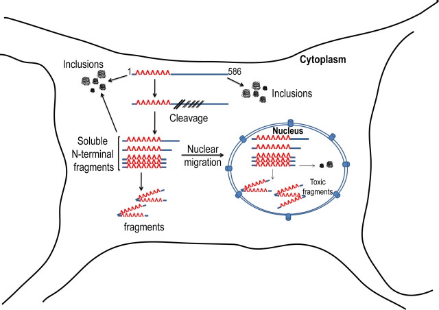
Huntington's disease (HD) is caused by a polyglutamine expansion in the Huntingtin (Htt) protein. Proteolytic cleavage of Htt into toxic N-terminal fragments is believed to be a key aspect of pathogenesis. The best characterized putative cleavage event is at amino acid 586, hypothesized to be mediated by caspase 6. A corollary of the caspase 6 cleavage hypothesis is that the caspase 6 fragment should be a toxic fragment. To test this hypothesis, and further characterize the role of this fragment, we have generated transgenic mice expressing the N-terminal 586 aa of Htt with a polyglutamine repeat length of 82 (N586-82Q), under the control of the prion promoter. N586-82Q mice show a clear progressive rotarod deficit by 4 months of age, and are hyperactive starting at 5 months, later changing to hypoactivity before early mortality. MRI studies reveal widespread brain atrophy, and histologic studies demonstrate an abundance of Htt aggregates, mostly cytoplasmic, which are predominantly composed of the N586-82Q polypeptide. Smaller soluble N-terminal fragments appear to accumulate over time, peaking at 4 months, and are predominantly found in the nuclear fraction. This model appears to have a phenotype more severe than current full-length Htt models, but less severe than HD mouse models expressing shorter Htt fragments. These studies suggest that the caspase 6 fragment may be a transient intermediate, that fragment size is a factor contributing to the rate of disease progression, and that short soluble nuclear fragments may be most relevant to pathogenesis.


Figures

Figure 1.
Figure 1.
Expression of the N586–82Q protein in three lines, and progressive fatal phenotype. A, Immunoblots of mouse brain homogenates showing the levels of N586–82Q transgene protein from three different mouse lines. Line 52 exhibits the highest level of expression. B, Regional distribution of N586–82Q from a 2-month-old transgenic line 52 mouse versus age-matched control. Top, N586–82Q is highly expressed throughout the brain. C, Representative photographs of wild-type mouse compared with N586–82Q line 52 at 9 months. N586–82Q mice are smaller, exhibit a hunched posture, and are less well groomed. Transgenic mice also exhibit limb clasping. D, Both lines begin to lose weight starting from ∼4 months and continue to lose weight over time. E, The average lifespan for each line falls approximately within 1 year.
Figure 2.
Figure 2.
N586–82Q mice exhibit a rotarod deficit, are initially hyperactive, and exhibit signs of cognitive impairment. A, B, Rotarod testing of two different lines demonstrates motor impairment. Four-month-old mice from line 52 (A) and line 91 (B) were compared to age-matched controls. For testing, the speed of the rod was set to 4 rpm to start and increased by 0.1 rpm every second. Mice received three trials per day for 3 consecutive days. Unpaired t test, *p < 0.05. Error bars represent SEs. C, D, Total activity of both lines was assessed by means of an open-field chamber for a period of 2 h during the dark phase of the cycle. Unpaired t test, ***p < 0.0001. Error bars represent SEs. E, Line 52 displays a cognitive deficit at 8 months. Similar results were obtained for line 91 at 10 months of age.
Figure 3.
Figure 3.
N586–82Q mice experience brain atrophy after an initial period of normal regional brain volumes. A, Sample images from an MRI scan using male mice from line 52, 3.5 months of age. B, C, Graphical representation of whole-brain atrophy and of specific brain regions following an MRI analysis. No significant change in brain volume was found at this time point. D, Sample images from an MRI scan using male mice from line 52, 9 months of age. E, F, Graphical representation of whole-brain atrophy and of specific brain regions following an MRI analysis. Most brain regions were affected. Most severely affected were cortex and hippocampus. Ventricles are enlarged (WT 3.1 mm2 vs HD 3.8 mm2). Unpaired t test, ***p < 0.0001. Error bars represent SEs.
Figure 4.
Figure 4.
N586–82Q is cleaved into smaller N-terminal fragments over time. A, The N586 fragment and the various antibody recognition sites. B, Whole-brain homogenates were prepared from male mice at the indicated ages. Full-length N586–82Q decreases over time, while smaller fragments accumulate. All antibodies used were capable of detecting N-terminal fragments with the exception of 115–129. The fastest-migrating fragment is smaller than amino acid 120.
Figure 5.
Figure 5.
Soluble N-terminal Htt fragments accumulate inside the nucleus. Whole-brain homogenates from mice aged 1 month (A) and 4 months (B) were subjected to nuclear and cytoplasmic extraction. A, B, Equal amounts of protein from each sample were run on a 4–12% Bis-Tris gel. The upper panels were probed with anti-Htt 81–90. A, N586–82Q can only be detected in the cytoplasm at 1 month of age. No N-terminal Htt fragments were detected at this time point. Similar results can be seen with anti-Htt 1–82 (lower panel). B, A greater amount of N-terminal Htt fragments can be seen in the nucleus than in the cytoplasm in mice aged 4 months. The next panel down shows an immunoblot probed with EM48, which was only capable of detecting full-length N586–82Q. Like 81–90, EM48 could only detect full-length N586–82Q in the cytoplasm. A, B, The last two panels are controls for fraction contamination. Neither fraction was contaminated.
Figure 6.
Figure 6.
Htt aggregates are composed of both N-terminal fragments and N586–82Q. A, Frozen/free-floating sections (30 mm) from 6-month-old N586–82Q mice were immunostained with EM48 (left panel) and anti-Htt 81–90 (right panel), followed by DAB and lightly counterstained with hematoxylin. Note that htt staining is absent from age-matched control. A high density of htt aggregates may be found throughout the cortex, striatum, and hippocampus. Representative images were taken from the cortex. The higher magnification (lower panel) shows EM48 to detect a large population of aggregates in the cytoplasm while anti-Htt 81–90 was capable of detecting more diffuse Htt, which appeared more nuclear. B, Free-floating sections from male mice, line 52 aged 1 year, were probed with the indicated antibodies. N-terminal Htt antibodies (1–82 and 81–90) were capable of detecting a multitude of Htt inclusion bodies. The more C-terminal anti-Htt antibody, 2166 (181–810), did not detect as many aggregates as either N-terminal antibodies but did appear to have more reactivity after the slices had been treated with formic acid (FA). Similarly, the N586 neo-epitope antibody, HD55, was also capable of detecting aggregates, and this reactivity was increased following FA treatment.
Figure 7.
Figure 7.
Htt aggregates are composed of both N-terminal fragments and N586–82Q with N586–82Q most abundant. A, Insoluble material from whole-brain homogenates from mice aged 4 months was treated with formic acid. Equal amounts of protein were loaded on a 4–12% Bis-Tris gel and immunoblots were probed with the indicated antibodies. Anti-Htt 2166 detects only N586–82Q. Immunoblots probed using either 3B5H10 and anti-polyQ antibody MW1 can detect both N586–82Q and N-terminal Htt fragments. Both antibodies indicate that N586–82Q is the major component of Htt aggregates. B, Insoluble material prepared from the nuclear and cytoplasmic fractionation was also subjected to formic acid treatment and immunoblotted using the same antibodies. All three antibodies used show N586–82Q to be the major component of cytoplasmic aggregates. Both 3B5H10 and MW1 show nuclear aggregates to be composed of only N-terminal Htt fragments. Asterisk indicates nonspecific band.
Figure 8.
Figure 8.
N586–82Q express substantially higher protein levels than N171–82Q and accumulate more of the “cp-1-like” fragment than YAC128. A, B, Immunoblots of mouse brain cortical homogenates aged 1 month showing the expression levels of N586–82Q relative to N171–82Q and full-length Htt in YAC128. A, Immunoreactivity using 81–90 demonstrated that the N586–82Q mice express much higher protein levels than the N171–82Q mice. Immunoblots using 1C2 and 1–82 revealed the same pattern of expression (data not shown). B, Immunoreactivity using the anti-polyglutamine; 1C2 revealed a similar expression level between N586–82Q and YAC128 mouse models. Immunoreactivity using anti-Htt 1–82 showed that protein expression levels were slightly higher in the N586–82Q mice. C, Immunoblots of brain homogenates prepared from cortices of both N586–82Q and YAC128 mice aged 4 months. Immunoreactivity using anti-Htt 81–90 revealed a greater accumulation of the fastest-migrating fragment, named here “cp-1-like” fragment in the N586–82Q mice compared to YAC128 mice. D, The ratio of fastest-migrating N-terminal fragment was obtained by comparing the amount of fragment generated from N586–82Q and full-length Htt in the respective mouse models. N586–82Q mice accumulated more of the “cp-1-like” fragment.
Figure 9.
Figure 9.
MRI studies reveal N586–82Q mice experience a greater loss in whole-brain volume earlier than full-length models. N586–82Q mice also die earlier than other full-length models. A, BACHD mice experience a 3.5% loss in brain volume at 18 months (n = 8). No significant change was seen before 18 months. YAC128 mice experience a 2% loss in total brain volume at 9 months, but this loss was not significant (NS). N586–82Q mice experience a 13.4% loss in whole-brain volume at 9 months of age. B, Summary of the survival of the various mouse models compared to N586–82Q. N586–82Q mice have an intermediate life span relative to full-length and other fragment models. Wavy line indicates no premature death.
Figure 10.
Figure 10.
Summary of N586–82Q proteolysis and aggregation pathway. The N586–82Q protein is subject to both proteolysis and aggregation inside the cytoplasm. Aggregates are abundantly cytoplasmic and are made up mostly of the N586–82Q protein. The N-terminal Htt fragments cleaved from N586–82Q in the cytoplasm can either remain soluble or form aggregates in the cytoplasm. Most of the N-terminal Htt fragments translocate to the nucleus, where they can also form aggregates.

References

    1. Aylward EH, Nopoulos PC, Ross CA, Langbehn DR, Pierson RK, Mills JA, Johnson HJ, Magnotta VA, Juhl AR, Paulsen JS. Longitudinal change in regional brain volumes in prodromal Huntington disease. J Neurol Neurosurg Psychiatry. 2011;82:405–410. - PMC - PubMed
    1. Cornett J, Cao F, Wang CE, Ross CA, Bates GP, Li SH, Li XJ. Polyglutamine expansion of huntingtin impairs its nuclear export. Nat Genet. 2005;37:198–204. - PubMed
    1. Crook ZR, Housman D. Huntington's disease: can mice lead the way to treatment? Neuron. 2011;69:423–435. - PMC - PubMed
    1. DiFiglia M, Sapp E, Chase KO, Davies SW, Bates GP, Vonsattel JP, Aronin N. Aggregation of huntingtin in neuronal intranuclear inclusions and dystrophic neurites in brain. Science. 1997;277:1990–1993. - PubMed
    1. Graham RK, Deng Y, Slow EJ, Haigh B, Bissada N, Lu G, Pearson J, Shehadeh J, Bertram L, Murphy Z, Warby SC, Doty CN, Roy S, Wellington CL, Leavitt BR, Raymond LA, Nicholson DW, Hayden MR. Cleavage at the caspase-6 site is required for neuronal dysfunction and degeneration due to mutant huntingtin. Cell. 2006;125:1179–1191. - PubMed

Publication types

MeSH terms