. 2012 Jan 4;32(1):322-30.
doi: 10.1523/JNEUROSCI.4070-11.2012.

Adenosine enhances sweet taste through A2B receptors in the taste bud

Affiliations

Adenosine enhances sweet taste through A2B receptors in the taste bud

Robin Dando et al. J Neurosci. .

Abstract

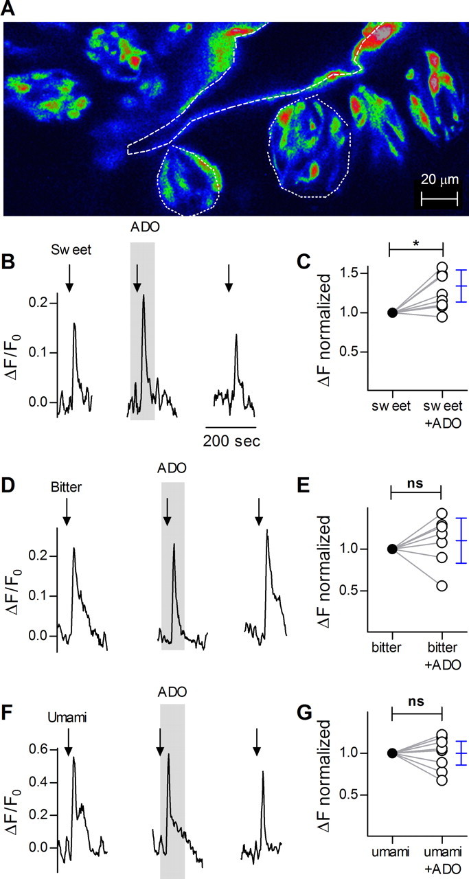
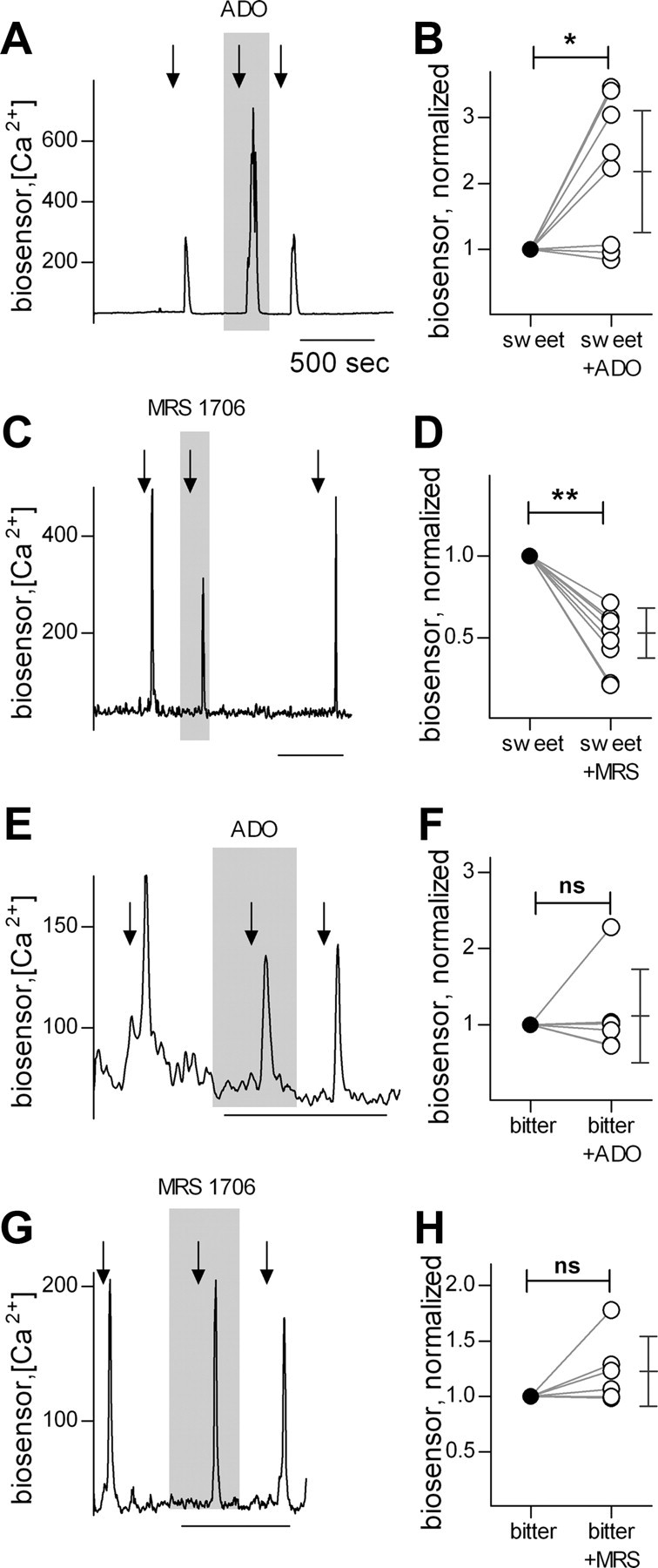
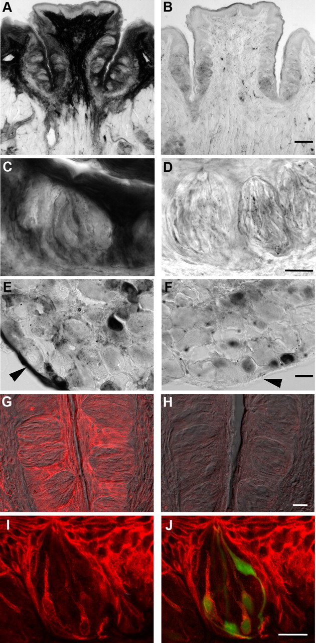
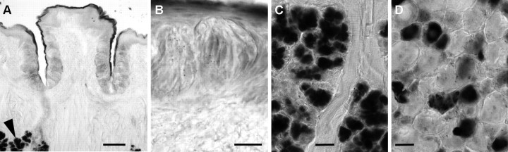
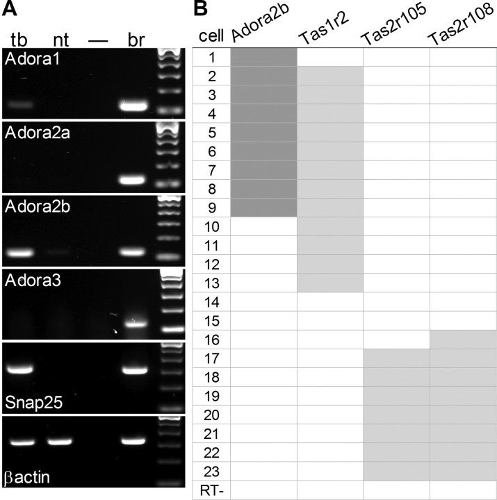
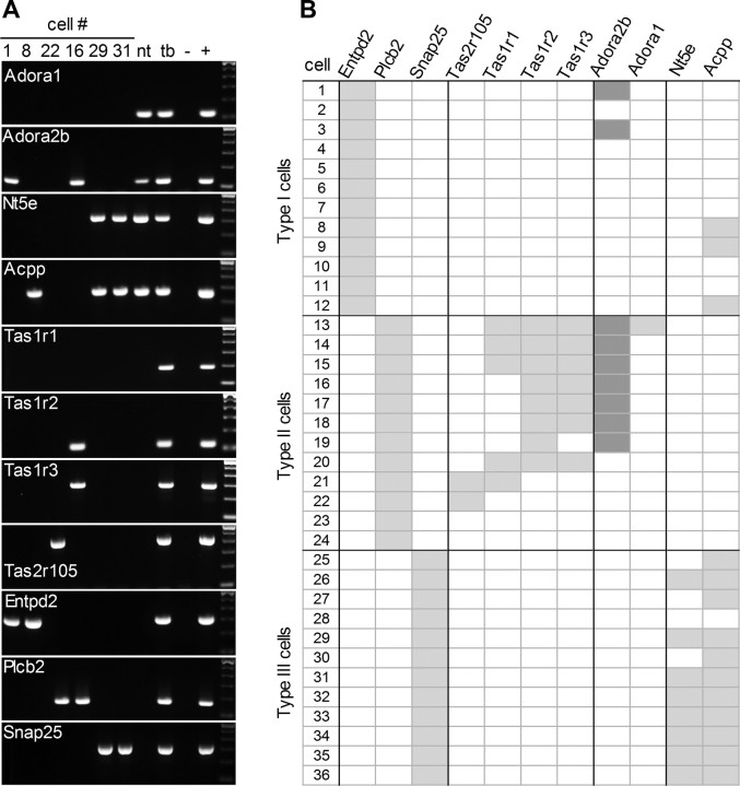
Mammalian taste buds use ATP as a neurotransmitter. Taste Receptor (type II) cells secrete ATP via gap junction hemichannels into the narrow extracellular spaces within a taste bud. This ATP excites primary sensory afferent fibers and also stimulates neighboring taste bud cells. Here we show that extracellular ATP is enzymatically degraded to adenosine within mouse vallate taste buds and that this nucleoside acts as an autocrine neuromodulator to selectively enhance sweet taste. In Receptor cells in a lingual slice preparation, Ca(2+) mobilization evoked by focally applied artificial sweeteners was significantly enhanced by adenosine (50 μM). Adenosine had no effect on bitter or umami taste responses, and the nucleoside did not affect Presynaptic (type III) taste cells. We also used biosensor cells to measure transmitter release from isolated taste buds. Adenosine (5 μM) enhanced ATP release evoked by sweet but not bitter taste stimuli. Using single-cell reverse transcriptase (RT)-PCR on isolated vallate taste cells, we show that many Receptor cells express the adenosine receptor, Adora2b, while Presynaptic (type III) and Glial-like (type I) cells seldom do. Furthermore, Adora2b receptors are significantly associated with expression of the sweet taste receptor subunit, Tas1r2. Adenosine is generated during taste stimulation mainly by the action of the ecto-5'-nucleotidase, NT5E, and to a lesser extent, prostatic acid phosphatase. Both these ecto-nucleotidases are expressed by Presynaptic cells, as shown by single-cell RT-PCR, enzyme histochemistry, and immunofluorescence. Our findings suggest that ATP released during taste reception is degraded to adenosine to exert positive modulation particularly on sweet taste.


Figures

Figure 1.
Figure 1.
Confocal imaging in lingual slices of taste cells responding to sweet, bitter and umami tastants. A, Micrograph of a lingual slice containing a vallate papilla, showing taste cells loaded with calcium green dextran (CaG dextran). Dashed line outlines the vallate crypt along which are embedded the taste buds (two examples outlined with dotted lines). B, Stimulation (arrows) of identified Receptor (type II) taste cell with mixture of sweet compounds SC45647 and saccharin, produces Ca2+ responses that are enhanced by the presence of adenosine (50 μm, present throughout the shaded area). Note that adenosine itself does not elicit a response. Ordinate, increase in CaG dextran fluorescence relative to baseline (see Materials and Methods). C, Summary of data from experiments as in B. The plot shows magnitude of responses to sweet stimulation before (filled circles) and during the presence of adenosine (open circles). Responses of each cell were normalized to its responses in the absence of adenosine. Student's t test, two tailed, repeated measures, p < 0.02. n = 8 cells. Blue symbols show the mean and 95% confidence interval. D, Ca2+ responses in vallate taste cells stimulated with a bitter taste mix (arrows) in absence or presence (shaded area) of adenosine as in B. E, Summary of results as in D, showing that adenosine does not affect bitter-evoked responses, p = 0.40, n = 7 cells. F, As in B, showing Ca2+ responses to umami stimulation. G, Summary of data showing that umami-evoked responses are unaffected by adenosine, p = 1.00, n = 9 cells. Calibrations in D and F as in B.
Figure 2.
Figure 2.
Taste-evoked transmitter (ATP) secretion from taste buds in response to sweet and bitter tastants, measured with biosensors. A, Traces show ATP biosensor responses evoked by stimulating the taste bud with a mixture of sweet compounds (arrows). Sweet-evoked ATP release is significantly enhanced by presence of adenosine in the bath (5 μm, shaded area). B, Summary of sweet-evoked ATP release. ATP biosensor responses were normalized to the response before adenosine. ATP release after adenosine was significantly elevated (p < 0.02, n = 8 cells, Student's paired two-tailed t test). Symbols on the right show the mean and 95% confidence interval. C, The A2B receptor antagonist, MRS 1706 (250 nm, present throughout shaded area), reduces sweet-evoked ATP release, indicating that sweet taste stimulation produces endogenous adenosine. Traces and stimuli as in A. D, Summary of data from experiments as in C (p < 0.01, n = 8 cells). E, Adenosine has no effect on bitter-evoked ATP release. F, Summary of bitter-evoked ATP release, as in E (p = 0.64, n = 6 cells). G, MRS 1706 has no effect on ATP release in response to bitter taste. H, Summary of data showing effects of MRS 1706 on bitter-evoked ATP release (p = 0.13, n = 6 cells). Calibrations in C, E, and G as in A.
Figure 3.
Figure 3.
Enzyme histochemical and immunofluorescent localization of ecto-5′-nucleotidase, NT5E. A, Cryosection of wild-type vallate papilla, developed to reveal enzymatic conversion of AMP to adenosine at pH 7.0. Very strong enzymatic activity, seen as dark staining, is present in the connective tissue core of the papilla, in non-taste epithelium on the lingual surface, and in keratinocytes surrounding taste buds. B, Cryosection of vallate papilla from a Nt5e−/− mouse, stained in parallel with that in A. Note the loss of prominent signal in the core of the papilla, in non-taste lingual epithelium, and in keratinocytes surrounding taste buds. C, At higher magnification, taste buds from wild-type mice can be seen to contain NT5E activity, particularly in slender cells. D, Higher magnification of taste buds from Nt5e−/− mouse. No distinct reaction product is seen in cells, although faint residual staining appears throughout the epithelium and taste buds. E, Dorsal root ganglion from wild-type mouse as in A and C shows that small- and medium-sized neurons, and the epineurium (arrowhead) of the ganglion, are stained for NT5E. F, Dorsal root ganglion from the same Nt5e−/− mouse as in B and D above shows that there is no staining in epineurium (arrowhead), but nucleotidase activity remains in small neurons, consistent with previous results (Sowa et al., 2010). G, Immunofluorescence for NT5E in vallate papilla of wild-type mouse, showing pronounced staining in keratinocytes surrounding the taste buds, and more modest staining in limited numbers of slender taste cells. Image is a merge of immunofluorescence (red) and Nomarski differential contrast optics. H, Sections of vallate papilla from Nt5e−/− mouse, stained in parallel and photographed with identical settings as in G. No signal is detected either in epithelium or in taste cells. I, J, Higher magnification of a vallate taste bud from a Gad1-GFP mouse stained for NT5E. The merged view (J) shows that NT5E signal (red) within the taste bud is in slender, GFP-positive cells (i.e., Type III /Presynaptic cells). Note how NT5E immunostaining (I, J) is similar to the shape and incidence of NT5E histochemical reaction product (C). Scale bars: A, D, 100 μm; B, E, 20 μm; C, F, 25 μm; G, H, 20 μm; I, J, 20 μm.
Figure 4.
Figure 4.
Enzyme histochemical detection of acid phosphatase activity. A, Cryosection of wild-type vallate papilla, developed to reveal enzymatic conversion of AMP to adenosine at pH 5.6. The enzymatic reaction conditions are optimized to detect ACPP. Only faint signal is detectable throughout the tissue with the exception of von Ebner glands located deep in the tissue (arrowhead). B, At higher magnification, reaction product is detected at low levels in the taste bud although individual cells cannot readily be distinguished. C, Von Ebner glands below taste epithelium show prominent signal in acini. D, Dorsal root ganglion from the same wild-type mouse as in A shows some small and medium sized neurons are positive for the acid phosphatase, while large neurons and the epineurium are negative. Scale bars: A, 100 μm; B, 20 μm; C, D, 25 μm.
Figure 5.
Figure 5.
Adenosine receptor Adora2b is associated with Tas1r2, a sweet taste receptor subunit in taste cells. A, RT-PCR on isolated vallate taste buds (tb) and non-taste lingual epithelium (nt) reveals prominent expression of Adora2b and lower levels of Adora1 in taste buds. Neither Adora2a nor Adora3 were detected. Positive and negative controls run in parallel included brain (br) and H2O in place of template (–). RT-PCR for Snap25 served as a positive control for taste buds. B, Single-cell RT-PCR on 23 individual GFP-positive cells (i.e., Type II Receptor cells) from taste buds of Plcb2-GFP mice. Aliquots (20%) of each cell's cDNA were tested for Adora2b, one sweet receptor subunit (Tas1r2) and two bitter receptors (Tas2r105, Tas2r108). One cell (RT−) was processed without reverse transcriptase, and yielded no PCR products.
Figure 6.
Figure 6.
Adora2b receptor is associated with taste cells that express the sweet taste receptor subunit, Tas1r2. T7-linear RNA amplification was performed on 36 individually isolated taste cells, followed by RT-PCR to classify cells into one of three cell types according to expression of the diagnostic mRNAs: Entpd2 (type I), Plcβ2 (type II) and Snap25 (type III). A, Two exemplar cells for each cell type are shown; each column contains data from a single cell. Each cell was also tested for expression of taste receptors, Tas2r105, Tas1r1, Tas1r2, and Tas1r3; of adenosine receptors, Adora1 and Adora2b; and of catabolic enzymes that produce adenosine, Nt5e and Acpp. To the right of individual cells are cDNAs from non-taste lingual epithelium (nt), isolated taste buds (tb), and a positive control (+) — either brain for the top 4 gels (adenosine receptors and enzymes), or vallate taste epithelium for the bottom 7 gels (taste receptors and diagnostic markers); parallel negative control reaction (−) without cDNA template. B, Aggregate data from all 36 taste cells. Adora2b is prominently detected in cells expressing Tas1r2 (i.e., sweet taste cells). Enzymes that convert AMP to adenosine (Nt5e and Acpp) were primarily in Presynaptic (type III) taste cells.

Similar articles

Cited by

References

    1. Baldwin SA, Beal PR, Yao SY, King AE, Cass CE, Young JD. The equilibrative nucleoside transporter family, SLC29. Pflugers Arch. 2004;447:735–743. - PubMed
    1. Bartel DL, Sullivan SL, Lavoie EG, Sévigny J, Finger TE. Nucleoside triphosphate diphosphohydrolase-2 is the ecto-ATPase of type I cells in taste buds. J Comp Neurol. 2006;497:1–12. - PMC - PubMed
    1. Caicedo A, Jafri MS, Roper SD. In situ Ca2+ imaging reveals neurotransmitter receptors for glutamate in taste receptor cells. J Neurosci. 2000;20:7978–7985. - PMC - PubMed
    1. Chandrashekar J, Mueller KL, Hoon MA, Adler E, Feng L, Guo W, Zuker CS, Ryba NJ. T2Rs function as bitter taste receptors. Cell. 2000;100:703–711. - PubMed
    1. Chattopadhyaya B, Di Cristo G, Higashiyama H, Knott GW, Kuhlman SJ, Welker E, Huang ZJ. Experience and activity-dependent maturation of perisomatic GABAergic innervation in primary visual cortex during a postnatal critical period. J Neurosci. 2004;24:9598–9611. - PMC - PubMed

Publication types

MeSH terms