Collision of three histologically distinct endometrial cancers of the uterus
- PMID: 22219620
- PMCID: PMC3247781
- DOI: 10.3346/jkms.2012.27.1.89
Collision of three histologically distinct endometrial cancers of the uterus
Abstract
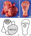
A collision tumor is defined by the presence of two separate masses in one organ, which are pathologically distinct. We described a 70-yr-old patient who complained of abnormal vaginal bleeding with a collision tumor of the uterine corpus. The patient received total hysterectomy, bilateral salphingo-oophorectomy, bilateral pelvic-paraaortic lymphadenectomy, omentectomy, and intraperitoneal chemotherapy. The uterine corpus revealed three separate masses, which were located at the fundus, anterior and posterior wall. Each tumor revealed three pathologically different components, which were malignant mixed müllerian tumor, papillary serous carcinoma, and endometrioid adenocarcinoma. Among these components, only the papillary serous carcinoma component invaded the underlying myometrium and metastasized to the regional lymph node. Adjuvant chemotherapy and radiation therapy were performed. The patient is still alive and has been healthy for the last 8 yr. We have reviewed previously reported cases of collision tumors which have occurred in the uterine corpus.
Keywords: Collision Tumor; Endometrial Neoplasms; Uterus.
Figures


References
-
- Lam KY, Khoo US, Cheung A. Collision of endometrioid carcinoma and stromal sarcoma of the uterus: a report of two cases. Int J Gynecol Pathol. 1999;18:77–81. - PubMed
-
- Gaertner EM, Farley JH, Taylor RR, Silver SA. Collision of uterine rhabdoid tumor and endometrioid adenocarcinoma: a case report and review of the literature. Int J Gynecol Pathol. 1999;18:396–401. - PubMed
-
- Lifschitz-Mercer B, Czernobilsky B, Dgani R, Dallenbach-Hellweg G, Moll R, Franke WW. Immunocytochemical study of an endometrial diffuse clear cell stromal sarcoma and other endometrial stromal sarcomas. Cancer. 1987;59:1494–1499. - PubMed
-
- Patwardhan JR, Gadgil RK. Collision tumour of the uterus. Indian J Cancer. 1969;6:194–197. - PubMed
Publication types
MeSH terms
Substances
LinkOut - more resources
Full Text Sources

