

High glucose may decrease the innate immune through TLRs in cornea epithelium
- PMID: 22219634
- PMCID: PMC3247169
High glucose may decrease the innate immune through TLRs in cornea epithelium
Abstract
Purpose: The purpose of this study was to investigate the high potential of glucose in inhibiting the innate immune in cultured human cornea epithelial cells (HCEC) and try to determine whether the role of high glucose on the HCEC relate to toll-like receptor (TLR)2 and TLR4.
Methods: Cells were cultured for 3 days in 5 mmol/l (normal glucose). Then high glucose (25 mmol/l) was added along with normal glucose with daily changes in media for 24 h. The cells were also treated with mannitol as an osmotic control. The cellular abundance of the mRNAs for TLR2 and TLR4 was determined by real-time polymerase chain reaction analysis. The proteins of TLR2 and TLR4 were also compared by immunofluorescent staining and western blot. The release of interleukin 6 (IL-6) and IL-8 from cultured HCEC was measured using enzyme-linked immunosorbent assays (ELISA) in the presence and absence of specific blocking antibodies to TLR2 and TLR4.
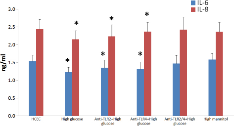
Results: Incubation of HCEC with high glucose showed that the mRNA expression of TLR2 and TLR4 was markedly inhibited. Immunofluorescent staining and western blot analysis confirmed that the protein expression of TLR2 and TLR4 was downregulated in response to high glucose. The result of ELISA also showed that the release of IL-6 and IL-8 can be inhibited by high glucose, but these inhibitions were partly counteracted after pretreatment with anti-TLR2 and/or anti-TLR4 monoclonal antibody. The results also showed that the osmotic control did not affect the expression of TLR2, TLR4, and IL-6, 8.
Conclusions: High glucose may decrease the innate immune through TLRs in cornea epithelium.
Figures


Similar articles
-
Glucocorticoids inhibit the innate immune system of human corneal fibroblast through their suppression of toll-like receptors.Mol Vis. 2009 Nov 20;15:2435-41. Mol Vis. 2009. PMID: 19956406 Free PMC article.
-
Toll-like receptors (TLRs) expression and function in response to inactivate hyphae of Fusarium solani in immortalized human corneal epithelial cells.Mol Vis. 2007 Oct 17;13:1953-61. Mol Vis. 2007. PMID: 17982419 Free PMC article.
-
Hydrocortisone suppression of the expression of VEGF may relate to toll-like receptor (TLR) 2 and 4.Curr Eye Res. 2009 Sep;34(9):777-84. doi: 10.1080/02713680903067919. Curr Eye Res. 2009. PMID: 19839871
-
[Aspergillus fumigatus activate human corneal epithelial cells via Toll-like receptor 2 and 4].Zhonghua Yan Ke Za Zhi. 2006 Jul;42(7):628-33. Zhonghua Yan Ke Za Zhi. 2006. PMID: 17081423 Chinese.
-
Thymic stromal lymphopoietin participates in the TLR2-and TLR4-dependent immune response triggered by Aspergillus fumigatus in human corneal cells.Exp Eye Res. 2021 Aug;209:108644. doi: 10.1016/j.exer.2021.108644. Epub 2021 May 31. Exp Eye Res. 2021. PMID: 34081998
Cited by
-
The Effect of Calcium and Glucose Concentration on Corneal Epithelial Cell Lines Differentiation, Proliferation, and Focal Adhesion Expression.Biores Open Access. 2019 Jun 5;8(1):74-83. doi: 10.1089/biores.2018.0036. eCollection 2019. Biores Open Access. 2019. PMID: 31179162 Free PMC article.
References
-
- Clark CM, Lee DA. Prevention and treatment of the complications of diabetes mellitus. N Engl J Med. 1995;332:1210–7. - PubMed
-
- Inoue K, Okugawa K, Amano S, Oshika T, Takamura E, Egami F, Umizu G, Aikawa K, Kato S. Blinking and superficial punctate keratopathy in patients with diabetes mellitus. Eye (Lond) 2005;19:418–21. - PubMed
-
- Rehany U, Ishii Y, Lahav M, Rumelt S. Ultrastructural changes in corneas of diabetic patients: an electron-microscopy study. Cornea. 2000;19:534–8. - PubMed
Publication types
MeSH terms
Substances
LinkOut - more resources
Full Text Sources