. 2011:17:3437-49.
Epub 2011 Dec 28.

Cornea lenticule viability and structural integrity after refractive lenticule extraction (ReLEx) and cryopreservation

Affiliations

Cornea lenticule viability and structural integrity after refractive lenticule extraction (ReLEx) and cryopreservation

Karim Mohamed-Noriega et al. Mol Vis. 2011.

Abstract

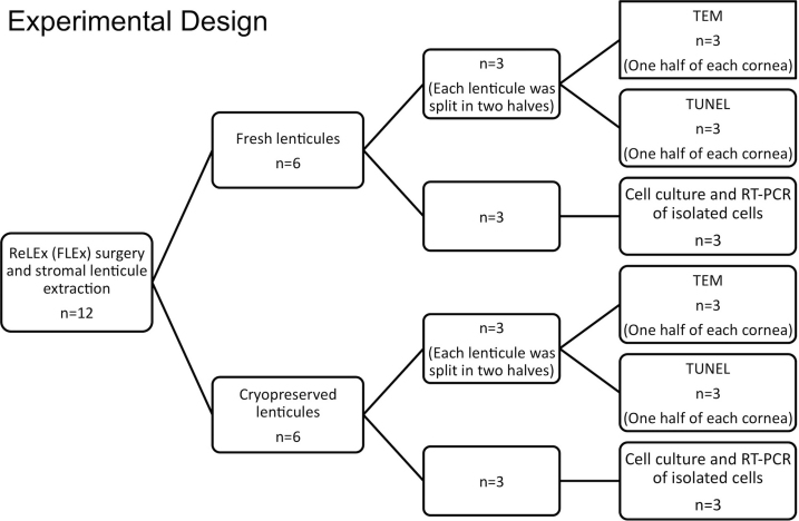
Purpose: To assess and compare keratocyte viability and collagen structure in cornea stroma lenticules collected immediately after refractive lenticule extraction (ReLEx) and one month after cryopreservation.

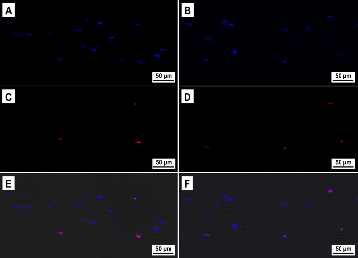
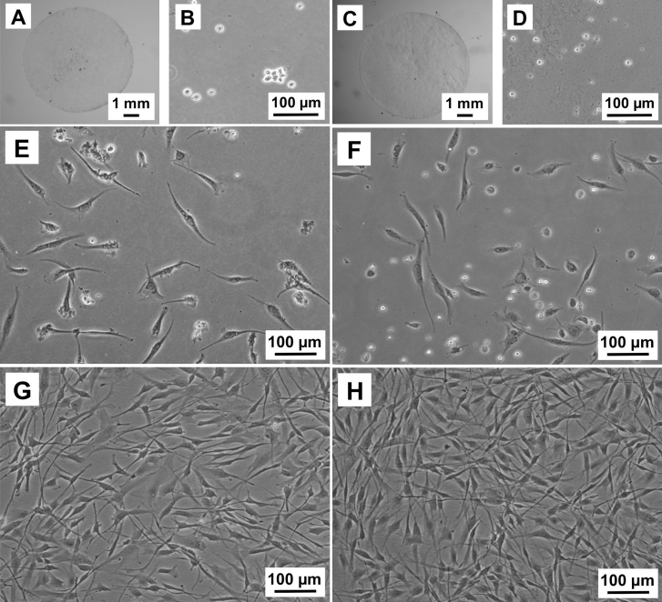
Methods: The fresh and cryopreserved human stroma lenticules procured after ReLEx were processed for ultrastructural analysis of keratocytes and collagen fibrils with transmission electron microscopy (TEM), apoptotic cell detection with deoxynucleotidyl transferase-mediated nick end labeling assay (TUNEL) assay, and cultured for keratocyte-specific gene expression analysis using reverse transcriptase polymerase chain reaction (RT-PCR).

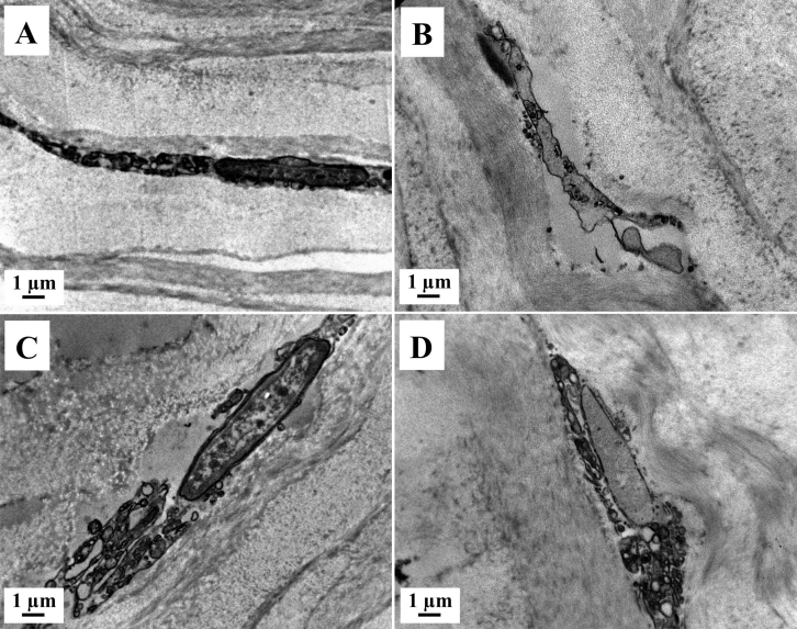
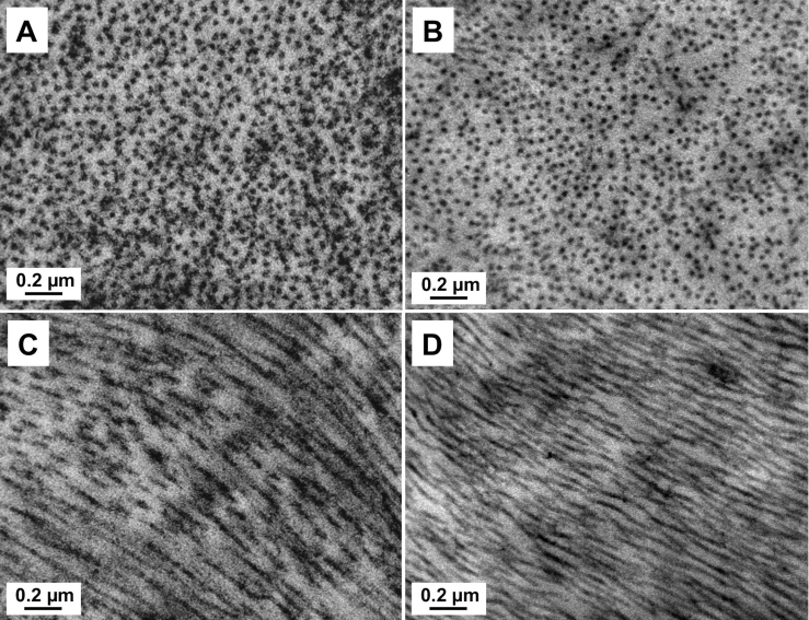
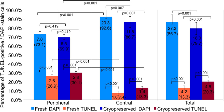
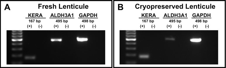
Results: The periphery of the lenticule had greater TUNEL-positive cells compared to the center of the lenticule in both fresh and cryopreserved groups. There was an increase in TUNEL-positive cells after cryopreservation, which was significantly higher in the center of the lenticule, but not in the periphery. TEM showed apoptotic, necrotic and viable quiescent keratocytes in fresh and cryopreserved lenticules. Collagen analysis with TEM showed a well preserved and well aligned structure in fresh and cryopreserved lenticules; without significant change in the total number of collagen fibrils but with an increased collagen fibril density (CFD) after cryopreservation. In vitro, isolated keratocytes derived from fresh and cryopreserved lenticules exhibited a typical fibroblastic phenotype. RT-PCR showed a positive gene expression for keratocan (KERA) and aldehyde dehydrogenase 3A1 (ALDH3A1) in cells isolated from fresh and cryopreserved lenticules.

Conclusions: The stromal lenticules extracted from ReLEx surgery remain viable after cryopreservation. Although they showed a decrease in CFD, the collagen architecture was preserved and there was good cellular viability.


Figures

Figure 1
Figure 1
Flow diagram showing the experimental design. ReLEx: Refractive lenticule extraction. FLEx: Femtosecond lenticule extraction. RT–PCR: reverse transcription-polymerase chain reaction. TEM: Transmission electron microscopy. TUNEL: deoxynucleotidyl transferase-mediated nick end labeling assay.
Figure 2
Figure 2
Laser scanning pattern and incisions created during femtosecond lenticule extraction (FLEx) procedure in the human cornea. A-D: Representative images of specific steps. A: Before the delivery of the laser the eye must be centered and full suction must be achieved. B: Creation of the posterior surface of the refractive lenticule with a scanning pattern in a centripetal direction (spiral in, arrow). C: Creation of the anterior surface of the refractive lenticule with a scanning pattern in a centrifugal direction (spiral out, arrow). D: Edge of the lenticule (long arrow) and creation of the periphery of the flap (short arrow).
Figure 3
Figure 3
Surgical steps in Femtosecond lenticule extraction (FLEx) A: Dissection, opening and lifting of the flap (arrow). B: Once the flap (short arrow) is flipped, the edge of the lenticule (long arrow) and the plane of the posterior surface of the lenticule are identified. C: The lenticule (arrow) is separated and removed from the cornea. D: After extraction of the lenticule, the previous location of the lenticule edge (arrow) can be identified in the surface of the stromal bed just before the flap is repositioned.
Figure 4
Figure 4
Transmission electron micrographs (TEM) of stromal lenticule showing keratocytes. A, C: Fresh lenticule. B, D: Cryopreserved lenticule. A, B: Apoptotic keratocytes with cromatin condensation and fragmentation, apoptotic bodies, loss of cytoplasm and cell shrinkage. C, D: Necrotic keratocyte, with incomplete nuclear membrane and vacuoles in the cytoplasm. Magnification, 8900×.
Figure 5
Figure 5
Transmission electron micrographs (TEM) of the stromal lenticule showing collagen fibrils. A, C: Fresh lenticule. B, D: Cryopreserved lenticule. A, B: Transversal section of collagen fibrils. C, D: Longitudinal section of collagen fibrils. Magnification, 50,000×.
Figure 6
Figure 6
TUNEL-positive (deoxynucleotidyl transferase-mediated nick end labeling assay) cells in fresh and cryopreserved human lenticules. A, C, E: Fresh samples. B, D, F: Cryopreserved samples. A, B: DAPI-stained (4',6-diamidino-2-phenylindole stain) cells. C, D: TUNEL-positive cells. E, F: Composite image of DAPI, TUNEL and Bright-field. Magnification, 200×.
Figure 7
Figure 7
Mean number (%) of TUNEL (deoxynucleotidyl transferase-mediated nick end labeling assay) positive cells and DAPI (4',6-diamidino-2-phenylindole) stain cells in fresh and cryopreserved lenticules extracted from a ReLEx (Refractive Lenticule Extraction) procedure. It was considered a significant difference when p<0.05.
Figure 8
Figure 8
Representative images of cultured keratocytes from ReLEx (Refractive Lenticule Extraction) lenticules. A, B, E, G: Fresh samples. C, D, F, H: Cryopreserved samples. A, C: ReLEx lenticule. B, D: Free floating stromal keratocytes following enzymatic digestion for at least 4 h in collagenase. E, F: Attached keratocytes beginning to elongate into spindle-like fibroblastic cells by Day 2 in culture. G, H: Confluent stromal fibroblasts after 7 days in culture.
Figure 9
Figure 9
Expression of keratocyte-specific markers in isolated cells from ReLEx (Refractive Lenticule Extraction) lenticules. Fresh (A) and cryopreserved (B) lenticues. Human keratocan (KERA) with 167 bp, aldehyde dehydrogenase 3A1 (ALDH3A1) with 495 bp and the housekeeping gene glyceraldehyde 3-phosphate dehydrogenase (GAPDH) with 498 bp. (+): Lenticule sample. (-): Negative control.

References

    1. Vaddavalli PK, Yoo SH. Femtosecond laser in-situ keratomileusis flap configurations. Curr Opin Ophthalmol. 2011;22:245–50. - PubMed
    1. Tran DB, Binder PS, Brame CL. LASIK flap revision using the IntraLase femtosecond laser. Int Ophthalmol Clin. 2008;48:51–63. - PubMed
    1. Kunert KS, Blum M, Duncker GIW, Sietmann R, Heichel J. Surface quality of human corneal lenticules after femtosecond laser surgery for myopia comparing different laser parameters. Graefes Arch Clin Exp Ophthalmol. 2011;249:1417–24. - PubMed
    1. Espandar L, Meyer J. Intraoperative and Postoperative Complications of Laser in situ Keratomileusis Flap Creation Using IntraLase Femtosecond Laser and Mechanical Microkeratomes. Middle East Afr J Ophthalmol. 2010;17:56–9. - PMC - PubMed
    1. Kim P, Sutton GL, Rootman DS. Applications of the femtosecond laser in corneal refractive surgery. Curr Opin Ophthalmol. 2011;22:238–44. - PubMed

Publication types

MeSH terms