Optimization of immunolocalization of cell cycle proteins in human corneal endothelial cells
- PMID: 22219645
- PMCID: PMC3249439
Optimization of immunolocalization of cell cycle proteins in human corneal endothelial cells
Abstract
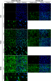
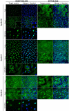
Purpose: En face observation of corneal endothelial cells (ECs) using flat-mounted whole corneas is theoretically much more informative than observation of cross-sections that show only a few cells. Nevertheless, it is not widespread for immunolocalization (IL) of proteins, probably because the endothelium, a superficial monolayer, behaves neither like a tissue in immunohistochemistry (IHC) nor like a cell culture in immunocytochemistry (ICC). In our study we optimized IL for ECs of flat-mounted human corneas to study the expression of cell cycle-related proteins.
Methods: We systematically screened 15 fixation and five antigen retrieval (AR) methods on 118 human fresh or stored corneas (organ culture at 31 °C), followed by conventional immunofluorescence labeling. First, in an attempt to define a universal protocol, we selected combinations able to correctly localize four proteins that are perfectly defined in ECs (zonula occludens-1 [ZO-1] and actin) or ubiquitous (heterogeneous nuclear ribonucleoprotein L [hnRNP L] and histone H3). Second, we screened protocols adapted to the revelation of 9 cell cycle proteins: Ki67, proliferating cell nuclear antigen (PCNA), minichromosome maintenance protein 2 (MCM2), cyclin D1, cyclin E, cyclin A, p16(Ink4a), p21(Cip1) and p27(Kip1). Primary antibody controls (positive controls) were performed on both epithelial cells of the same, simultaneously-stained whole corneas, and by ICC on human ECs in in vitro non-confluent cultures. Both controls are known to contain proliferating cells. IL efficiency was evaluated by two observers in a masked fashion. Correct localization at optical microscopy level in ECs was define as clear labeling with no background, homogeneous staining, agreement with previous works on ECs and/or protein functions, as well as a meaningful IL in proliferating cells of both controls.
Results: The common fixation with 4% formaldehyde (gold standard for IHC) failed to reveal 12 of the 13 proteins. In contrast, they were all revealed using either 0.5% formaldehyde at room temperature (RT) during 30 min alone or followed by AR with sodium dodecyl sulfate or trypsin, or pure methanol for 30 min at RT. Individual optimization was nevertheless often required to optimize the labeling. Ki67 was absent in both fresh and stored corneas, whereas PCNA was found in the nucleus, and MCM2 in the cytoplasm, of all ECs. Cyclin D1 was found in the cytoplasm in a paranuclear pattern much more visible after corneal storage. Cyclin E and cyclin A were respectively nuclear and cytoplasmic, unmodified by storage. P21 was not found in ECs with three different antibodies. P16 and p27 were exclusively nuclear, unmodified by storage.
Conclusions: IL in ECs of flat-mounted whole human corneas requires a specific sample preparation, especially to avoid overfixation with aldehydes that probably easily masks epitopes. En face observation allows easy analysis of labeling pattern within the endothelial layer and clear subcellular localization, neither of which had previously been described for PCNA, MCM2, or cyclin D1.
Figures


Similar articles
-
Optimization of immunostaining on flat-mounted human corneas.Mol Vis. 2015 Dec 30;21:1345-56. eCollection 2015. Mol Vis. 2015. PMID: 26788027 Free PMC article.
-
Expression of senescence-related genes in human corneal endothelial cells.Mol Vis. 2008 Jan 29;14:161-70. Mol Vis. 2008. PMID: 18334933 Free PMC article.
-
Expression patterns of cyclins D1, E and cyclin-dependent kinase inhibitors p21(Waf1/Cip1) and p27(Kip1) in urothelial carcinoma: correlation with other cell-cycle-related proteins (Rb, p53, Ki-67 and PCNA) and clinicopathological features.Urol Int. 2004;73(1):65-73. doi: 10.1159/000078807. Urol Int. 2004. PMID: 15263796
-
p27kip1 Antisense-induced proliferative activity of rat corneal endothelial cells.Invest Ophthalmol Vis Sci. 2004 Jun;45(6):1763-70. doi: 10.1167/iovs.03-0885. Invest Ophthalmol Vis Sci. 2004. PMID: 15161838
-
Involvement of p27KIP1 in the proliferation of the developing corneal endothelium.Invest Ophthalmol Vis Sci. 2004 Jul;45(7):2163-7. doi: 10.1167/iovs.03-1238. Invest Ophthalmol Vis Sci. 2004. PMID: 15223790
Cited by
-
Collagen Substrate Stiffness Anisotropy Affects Cellular Elongation, Nuclear Shape, and Stem Cell Fate toward Anisotropic Tissue Lineage.Adv Healthc Mater. 2016 Sep;5(17):2237-47. doi: 10.1002/adhm.201600284. Epub 2016 Jul 5. Adv Healthc Mater. 2016. PMID: 27377355 Free PMC article.
-
Investigating the Role of TGF-β Signaling Pathways in Human Corneal Endothelial Cell Primary Culture.Cells. 2023 Jun 14;12(12):1624. doi: 10.3390/cells12121624. Cells. 2023. PMID: 37371094 Free PMC article.
-
Investigating the role of molecular coating in human corneal endothelial cell primary culture using artificial intelligence-driven image analysis.Sci Rep. 2025 Aug 25;15(1):31301. doi: 10.1038/s41598-025-14367-4. Sci Rep. 2025. PMID: 40854928 Free PMC article.
-
Technique of flat-mount immunostaining for mapping the olfactory epithelium and counting the olfactory sensory neurons.PLoS One. 2023 Jan 17;18(1):e0280497. doi: 10.1371/journal.pone.0280497. eCollection 2023. PLoS One. 2023. PMID: 36649285 Free PMC article.
-
In Vivo Labeling and Tracking of Proliferating Corneal Endothelial Cells by 5-Ethynyl-2'-Deoxyuridine in Rabbits.Transl Vis Sci Technol. 2021 Sep 1;10(11):7. doi: 10.1167/tvst.10.11.7. Transl Vis Sci Technol. 2021. PMID: 34478491 Free PMC article.
References
-
- Murphy C, Alvarado J, Juster R, Maglio M. Prenatal and postnatal cellularity of the human corneal endothelium. A quantitative histologic study. Invest Ophthalmol Vis Sci. 1984;25:312–22. - PubMed
-
- Bourne WM, Nelson LR, Hodge DO. Central corneal endothelial cell changes over a ten-year period. Invest Ophthalmol Vis Sci. 1997;38:779–82. - PubMed
-
- Joyce NC. Proliferative capacity of the corneal endothelium. Prog Retin Eye Res. 2003;22:359–89. - PubMed
-
- Wilson SE, Weng J, Blair S, He YG, Lloyd S. Expression of E6/E7 or SV40 large T antigen-coding oncogenes in human corneal endothelial cells indicates regulated high-proliferative capacity. Invest Ophthalmol Vis Sci. 1995;36:32–40. - PubMed
Publication types
MeSH terms
Substances
LinkOut - more resources
Full Text Sources
Research Materials
Miscellaneous
