. 2010;10(6):5724-57.
doi: 10.3390/s100605724. Epub 2010 Jun 8.

Windows on the human body--in vivo high-field magnetic resonance research and applications in medicine and psychology

Affiliations

Windows on the human body--in vivo high-field magnetic resonance research and applications in medicine and psychology

Ewald Moser et al. Sensors (Basel). 2010.

Abstract

Analogous to the evolution of biological sensor-systems, the progress in "medical sensor-systems", i.e., diagnostic procedures, is paradigmatically described. Outstanding highlights of this progress are magnetic resonance imaging (MRI) and spectroscopy (MRS), which enable non-invasive, in vivo acquisition of morphological, functional, and metabolic information from the human body with unsurpassed quality. Recent achievements in high and ultra-high field MR (at 3 and 7 Tesla) are described, and representative research applications in Medicine and Psychology in Austria are discussed. Finally, an overview of current and prospective research in multi-modal imaging, potential clinical applications, as well as current limitations and challenges is given.

Keywords: 1H; 23Na; 31P; EEG; MRI; MRS; SCP; SWI; brain; cartilage; fMRI; joints; magnetic field strength; magnetic resonance; multi modal imaging; sensors; skeletal muscle.


Figures

Figure 1.
Figure 1.
Comparison of T1 and T2 contrast at 3 T (12-channel head coil): T1-weighted, transverse slice through the brain (left). As can be seen T1-weighted images provide an excellent contrast between gray and white matter (black arrow) as well as a good definition of the ventricles. On the right side the same slice with T2-weighted contrast is shown; note the lower gray and white matter contrast but the excellent definition of the ventricles (white arrow).
Figure 2.
Figure 2.
Susceptibility-Weighted Imaging at 7 T (8-channel head coil); this image represents a minimum intensity projection over a 6 mm slab. Note that both, veins and iron containing structures like the basal ganglia appear hypo intense. This image was acquired using an echo time of 15 ms and a resolution of 0.3 × 0.3 × 1.2 mm.
Figure 3.
Figure 3.
7 Tesla gradient-echo image of the knee joint in the coronal plane. The cartilage layers and the menisci are shown in high resolution and exquisite detail and will help to detect even subtle pathologies of these structures. Additionally, the trabecular structure of the bones is shown in ultra-high resolution, which may promote osteoporosis research.
Figure 4.
Figure 4.
The 7 Tesla sodium image in the sagittal plane shows cartilage with high sodium content in the lateral femoral tibial joint cavity of the knee joint. Since sodium content correlates with the proteoglycan content of cartilage, which is related to the biomechanical properties of cartilage, a sodium image of cartilage provides biomechanical information. Note that even the thin cartilage layers of the proximal tibio-fibular joint are shown with sodium imaging.
Figure 5.
Figure 5.
Brain regions that are activated by the different, ultra-short presentation durations using a linear regressor comprising the presentation durations of 1, 3, 5, 7, 9, and 11 ms. The sagittal map shows significant BOLD-signal increases projected onto the anatomical brain image of the subject (p < 0.05 FWE-corrected).
Figure 6.
Figure 6.
Activations associated with subjective perception. For this analysis a sigmoidal regressor corresponding to the probability with which the faces’ gender was detected (A) was used. The sagittal map (B) shows significant BOLD-signal increases projected onto the anatomical brain image of the particular subject (p < 0.05 FWE-corrected).
Figure 6.
Figure 6.
Activations associated with subjective perception. For this analysis a sigmoidal regressor corresponding to the probability with which the faces’ gender was detected (A) was used. The sagittal map (B) shows significant BOLD-signal increases projected onto the anatomical brain image of the particular subject (p < 0.05 FWE-corrected).
Figure 7.
Figure 7.
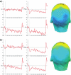
Averaged waveforms at representative scalp locations (left and middle row) and scalp potential topographies (right; dark blue color indicates stronger activation) averaged across the interval from 2,000–5,000 ms post stimulus of a single subject. (a) SCPs recorded inside the scanner without fMRI acquisition, (b) SCPs recorded while acquiring fMRI data.
Figure 8.
Figure 8.
SCP-based source localization within the interval 2,000—5,000 ms post stimulus of a single subject. (a) SCPs recorded inside the scanner without fMRI acquisition, (b) SCPs recorded while acquiring fMRI data.
Figure 9.
Figure 9.
Single-subject fMRI results of the visual stimulation paradigm as used in Figures 7b and 8b (p < 0.001, uncorr.).
Figure 10.
Figure 10.
1H spectra of skeletal muscle acquired at 3 T (a) and 7 T (b), demonstrating improvement of spectral quality with increasing B0 field strength. Advantages of higher B0 field strength are improved separation of resonances and higher signal to noise ratio (b).
Figure 11.
Figure 11.
Dynamic localized 31P MRS of skeletal muscle acquired at 3 Tesla (left) and 7 Tesla (right). Signal gain at 7 T (in connection with an improved acquisition method, 31P semi-LASER) allows higher time resolution and yet surpasses spectral quality for quantification of creatine phosphate (PCr) and inorganic phosphate [87] time courses during and after aerobic exercise of human muscle, compared to a similar measurement at 3 T. Spectra were scaled to equal noise level.
Figure 12.
Figure 12.
High field whole body MR scanner Siemens TIM Trio (3 Tesla, left) with a 12-channel receive-only head coil (Siemens). A cylindrically polarized (CP) whole body RF coil for transmitting 123 MHz (1H at 3 T) radio frequency signals and B0 gradient coils are installed in the scanner bore. Various Rx or local Tx/Rx coils can be used, e.g., a 32-channel receive head coil also by Siemens (right).
Figure 13.
Figure 13.
Ultra-high field whole body MR scanner Siemens Magnetom 7 T (left) with 24-channel (transmit-) receive coil (by Nova Medical) placed on the patient bed. B0 gradient-coils are installed in the scanner bore. The 24-channel Rx head coil (top right) is equipped with a transmit coil (outer cylinder), tuned to 300 MHz (Larmor frequency of 1H at 7 T). Single-loop transmit/receive surface coil by RAPID Biomedical (bottom right).

References

    1. Moser E. Ultra-high-field Magnetic Resonance: why and when? World J. Radiol. 2010;2:37–40. - PMC - PubMed
    1. Ugurbil K., Adriany G., Andersen P., Chen W., Garwood M., Gruetter R., Henry P.G., Kim S.G., Lieu H., Tkac I., Vaughan T., van De Moortele P.F., Yacoub E., Zhu X.H. Ultrahigh field magnetic resonance imaging and spectroscopy. Magn. Reson. Imaging. 2003;21:1263–1281. - PubMed
    1. De Graaf RA. In Vivo NMR Spectroscopy: Principles and Techniques. Wiley; Chichester, UK; New York, NY, USA: 1998. p. xxi.p. 508.
    1. Haacke E.M. Magnetic resonance imaging: physical principles and sequence design. Wiley & Sons; New York, NY, USA: 1999. p. xxvii.p. 914.
    1. Filippi M. fMRI Techniques and Protocols. Springer; New York, NY, USA: 2009. p. xiii.p. 843.

Publication types

MeSH terms