

Esophageal squamous cell carcinoma with marked eosinophil infiltration
- PMID: 22220139
- PMCID: PMC3250651
- DOI: 10.1159/000332441
Esophageal squamous cell carcinoma with marked eosinophil infiltration
Abstract
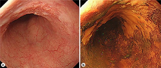
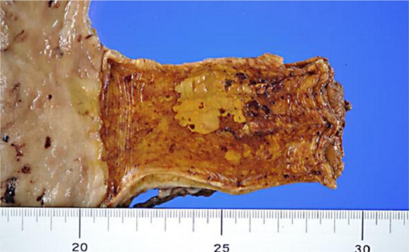
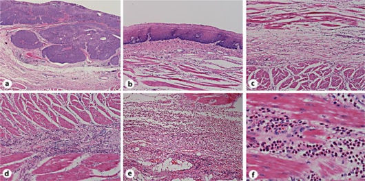
We report a case of esophageal squamous cell carcinoma (SCC) with marked eosinophil infiltration which was identified postoperatively in the esophageal wall in areas not surrounding the SCC. The eosinophil infiltration was seen in the submucosa, muscle and adventitia, but not in the mucosa. Eosinophilic esophagitis (EoE) is a pathological condition defined as eosinophil infiltration within the esophageal mucosa. Eosinophil infiltration at the invasion front of esophageal SCC is termed tumor-associated tissue eosinophilia (TATE). However, the eosinophil infiltration in this case may be pathologically different from both EoE and TATE. To our knowledge, this is the first report of esophageal SCC with eosinophil infiltration.
Keywords: Eosinophil infiltration; Esophageal squamous cell carcinoma; Esophagectomy.
Figures
Similar articles
-
Normalized serum eosinophil peroxidase levels are inversely correlated with esophageal eosinophilia in eosinophilic esophagitis.Dis Esophagus. 2018 Feb 1;31(2):dox139. doi: 10.1093/dote/dox139. Dis Esophagus. 2018. PMID: 29228243 Free PMC article.
-
Integrin αM activation and upregulation on esophageal eosinophils and periostin-mediated eosinophil survival in eosinophilic esophagitis.Immunol Cell Biol. 2018 Apr;96(4):426-438. doi: 10.1111/imcb.12018. Epub 2018 Mar 6. Immunol Cell Biol. 2018. PMID: 29424023
-
A new eosinophilic esophagitis (EoE)-like disease without tissue eosinophilia found in EoE families.Allergy. 2016 Jun;71(6):889-900. doi: 10.1111/all.12879. Epub 2016 Apr 1. Allergy. 2016. PMID: 26970242
-
Eosinophilic esophagitis: asthma of the esophagus?Dig Dis. 2014;32(1-2):54-60. doi: 10.1159/000357010. Epub 2014 Feb 28. Dig Dis. 2014. PMID: 24603381 Review.
-
[Eosinophilic esophagitis in children: analysis of 22 cases].Zhonghua Er Ke Za Zhi. 2017 Jul 2;55(7):499-503. doi: 10.3760/cma.j.issn.0578-1310.2017.07.006. Zhonghua Er Ke Za Zhi. 2017. PMID: 28728257 Review. Chinese.
Cited by
-
Symptom-based diagnostic approach for eosinophilic esophagitis.J Gastroenterol. 2020 Sep;55(9):833-845. doi: 10.1007/s00535-020-01701-y. Epub 2020 Jul 27. J Gastroenterol. 2020. PMID: 32720208 Free PMC article. Review.
-
Clinical impact of tumor-infiltrating inflammatory cells in primary small cell esophageal carcinoma.Int J Mol Sci. 2014 May 30;15(6):9718-34. doi: 10.3390/ijms15069718. Int J Mol Sci. 2014. PMID: 24886814 Free PMC article.
-
Innate Immune Cells in the Esophageal Tumor Microenvironment.Front Immunol. 2021 Apr 28;12:654731. doi: 10.3389/fimmu.2021.654731. eCollection 2021. Front Immunol. 2021. PMID: 33995371 Free PMC article. Review.
References
-
- Liacouras CA, Markowitz JE. Eosinophilic esophagitis: a subset of eosinophilic gastroenteritis. Curr Gastroenterol Rep. 1999;1:253–258. - PubMed
-
- Klein NC, Hargrove RL, Sleisenger MH, Jeffries GH. Eosinophilic gastroenteritis. Medicine. 1970;49:299–319. - PubMed
-
- Rothenberg ME, Hogan SP. The eosinophil. Annu Rev Immunol. 2006;24:147–174. - PubMed
-
- Furuta K, Adachi K, Kowari K, Mishima Y, Imaoka H, Kadota C, Koshino K, Miyake T, Kadowaki Y, Furuta K, Kazumori H, Sato S, Ishihara S, Amano Y, Honda M, Kinoshita Y. A Japanese case of eosinophilic esophagitis. J Gastroenterol. 2006;41:706–710. - PubMed
Publication types
LinkOut - more resources
Full Text Sources
Research Materials
