. 2012 Mar-Apr;88(2):461-8.
doi: 10.1111/j.1751-1097.2012.01081.x. Epub 2012 Jan 25.

Lysosomal signaling enhances mitochondria-mediated photodynamic therapy in A431 cancer cells: role of iron

Affiliations

Lysosomal signaling enhances mitochondria-mediated photodynamic therapy in A431 cancer cells: role of iron

Shalini Saggu et al. Photochem Photobiol. 2012 Mar-Apr.

Abstract

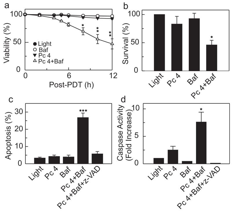
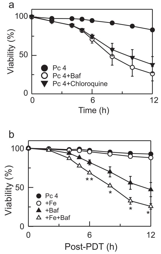
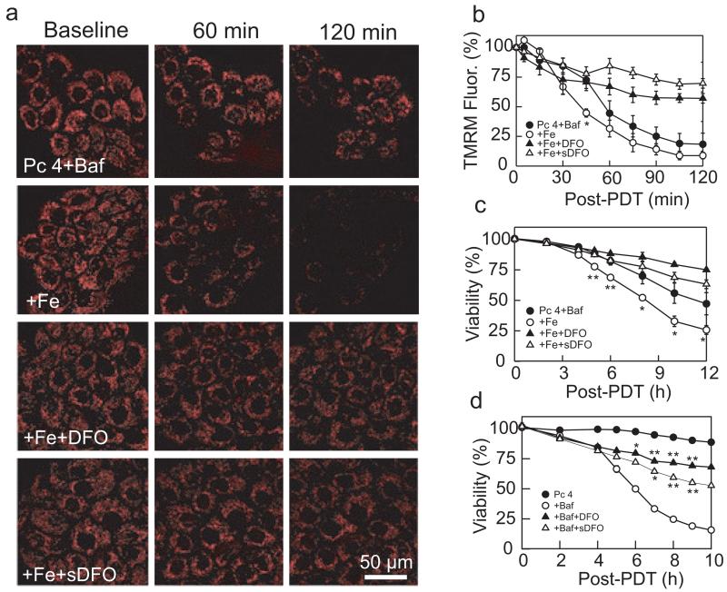
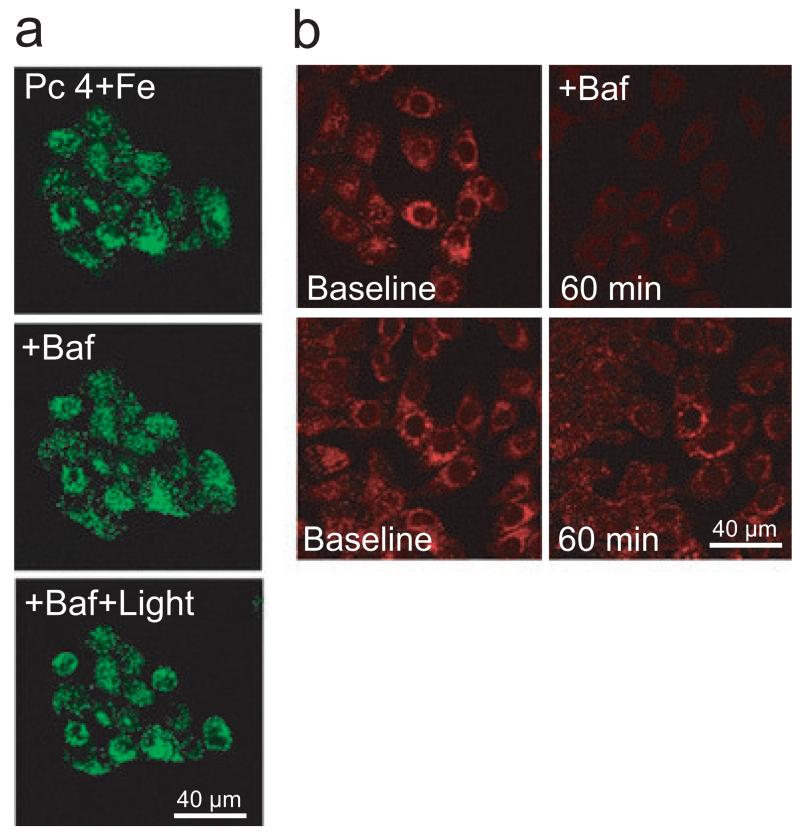
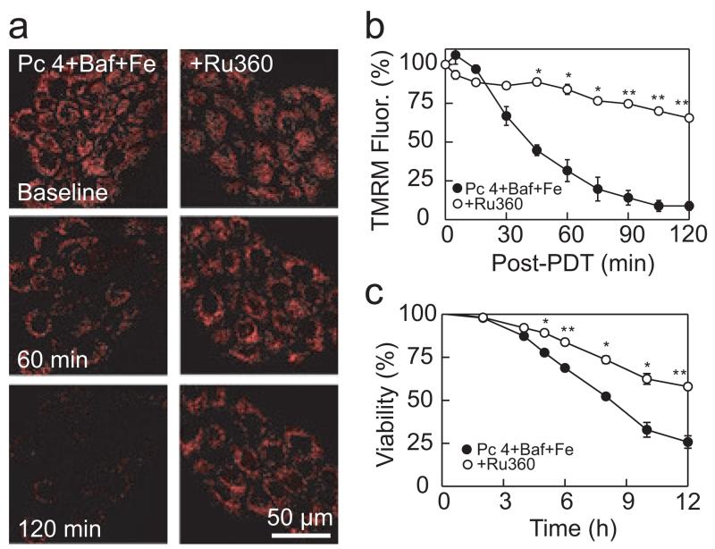
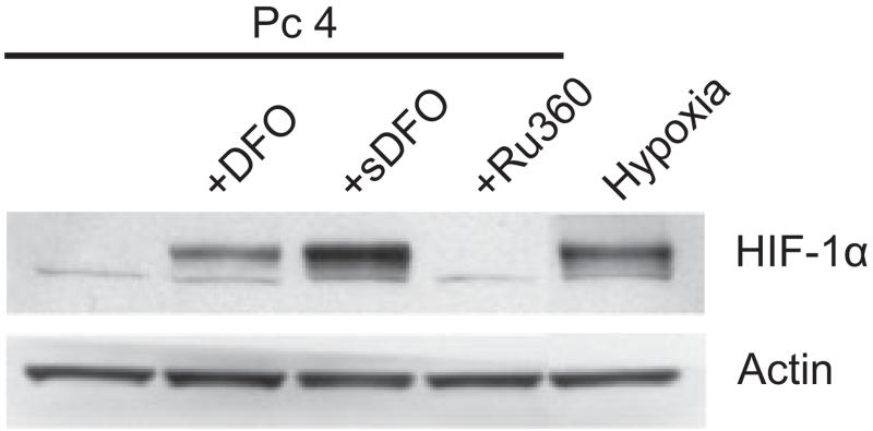
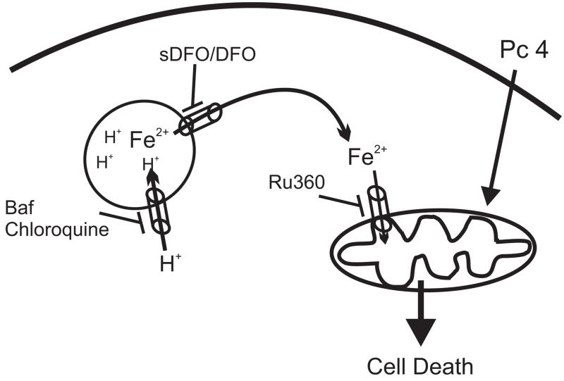
In photodynamic therapy (PDT), light activates a photosensitizer added to a tissue, resulting in singlet oxygen formation and cell death. The photosensitizer phthalocyanine 4 (Pc 4) localizes primarily to mitochondrial membranes in cancer cells, resulting in mitochondria-mediated cell death. The aim of this study was to determine how lysosomes contribute to PDT-induced cell killing by mitochondria-targeted photosensitizers such as Pc 4. We monitored cell killing of A431 cells after Pc 4-PDT in the presence and absence of bafilomycin, an inhibitor of the vacuolar proton pump of lysosomes and endosomes. Bafilomycin was not toxic by itself, but greatly enhanced Pc 4-PDT-induced cell killing. To investigate whether iron loading of lysosomes affects bafilomycin-induced killing, cells were incubated with ammonium ferric citrate (30 μM) for 30 h prior to PDT. Ammonium ferric citrate enhanced Pc 4 plus bafilomycin-induced cell killing without having toxicity by itself. Iron chelators (desferrioxamine and starch-desferrioxamine) and the inhibitor of the mitochondrial calcium (and ferrous iron) uniporter, Ru360, protected against Pc 4 plus bafilomycin toxicity. These results support the conclusion that chelatable iron stored in the lysosomes enhances the efficacy of bafilomycin-mediated PDT and that lysosomal disruption augments PDT with Pc 4.


Figures

Figure 1
Figure 1
Bafilomycin enhances Pc 4-PDT-mediated cell death. A431 cells were plated on 96-well plates (6 000 cells per well) and cultured for 24 h. Subsequently, cells were incubated with Pc 4 (25 nm) in FBS containing culture medium. After 18 h, medium was changed to fresh medium supplemented with Insulin-Transferrin-Selenium-X Reagent and PI (30 μm), but omitting FBS, followed by incubation with bafilomycin (50 nm; Baf) for 1 h, where indicated. Subsequently, cells were exposed to light as described in the Materials and Methods section. (a) Viability was assessed by PI exclusion using fluorometry. Results are expressed as percent viability at 0 h. Data represent three independent experiments (mean ± SEM) performed in quadruplicate. *P < 0.05; **P < 0.01 compared with Pc 4. (b) Cells (330 000 cells/6-cm Petri dish) were treated and irradiated as in (a). Subsequently, cells were trypsinized and plated on 6-cm Petri dishes. After 14 days, colonies were stained with crystal violet and counted. Results are expressed as percent colonies of light-treated cultures. Data represent three or more independent experiments performed in triplicate. *P < 0.05 compared with Pc 4 (one-tailed t-test). (c) Cells were plated on 96-well plates (6 000 cells per well) and treated as described in (a). z-VAD (10 μm) was added, as indicated, 1 h prior to irradiation. Four hours after irradiation, apoptotic nuclei were scored with a fluorescence microscope as described in the Materials and Methods section. At least 200 cells were counted from three different microscopic fields for each treatment group. Results are expressed as percent apoptotic nuclei. Data represent three independent experiments (mean ± SEM) performed in triplicate. ***P < 0.005 compared with Pc 4. (d) Cells were plated on 6-well plates (120 000 cells per well) and treated as described in (a). Four hours after irradiation, cell lysates were prepared as described in the Materials and Methods section. Caspase 3/7 activity was normalized for protein content, and results are expressed as fold increase from light-treated cells. Data represent three independent experiments (mean ± SEM) performed in triplicate. *P < 0.05 compared with Pc 4 (one-tailed t-test).
Figure 2
Figure 2
Chloroquine enhances Pc 4-PDT-induced cell killing. (a) Cells were treated as described in Fig. 1a. As indicated, 50 nm bafilomycin or 50 μm chloroquine was added 1 h before irradiation, and cell viability was monitored with PI fluorometry. Results are expressed as percent viability of 0 h. Data represent three independent experiments performed in quadruplicate. (b) Cells were cultured in medium containing 30 μm Fe3+ for 24 h, as indicated. Subsequently, Fe3+ was washed out and cells were incubated with 25 nm Pc 4 for 18 h. Medium was replaced with medium supplemented with Insulin-Transferrin-Selenium-X Reagent and omitting FBS. Bafilomycin was added, as indicated. Cell viability was monitored with PI fluorometry. Data represent three independent experiments (mean ± SEM) performed in quadruplicate. *P < 0.05; **P < 0.01 compared with Pc 4 + Baf.
Figure 3
Figure 3
Bafilomycin enhances Pc 4-PDT-mediated mitochondrial depolarization. (a) Cells were cultured on glass-bottomed Petri dishes in the presence and absence of Fe3+(30 μm), as indicated. Subsequently, cells were incubated with Pc 4 (25 nm) in the presence and absence of DFO (1 mm) or sDFO for 18 h. Cells were loaded with 250 nm TMRM and subsequently incubated with TMRM (50 nm) and bafilomycin (Baf, 50 nm) for 1 h before irradiation. Red fluorescence of TMRM was imaged with laser scanning confocal microscopy before (0 min) and at 60 and 120 min after irradiation. Note loss of TMRM fluorescence after Pc 4-PDT in presence of Baf + Fe, which was prevented by DFO and sDFO. Representative images from three independent experiments. (b) average TMRM fluorescence after background subtraction under conditions described in panel (a) was determined every 15 min for 120 min. Results are expressed as percent TMRM fluorescence of 0 min. Data are means calculated from analyses of 82–100 cells per treatment group obtained from three independent experiments (mean ± SEM). *P < 0.05 compared with Pc 4 + Baf. (c) Cells were plated on 96-well plates and treated under same conditions as in panel (a). Viability was monitored using PI fluorometry. Results are expressed as percent viability of 0 min. Data represent three independent experiments (mean ± SEM) performed in quadruplicate. *P < 0.05; **P < 0.01 compared with Pc 4 + Baf. (d) Cells were exposed to Pc 4 (50 nm), Pc 4-PDT and Pc 4-PDT + Baf in the presence and absence of DFO and sDFO. *P < 0.05; **P < 0.01 compared to Pc 4 + Baf.
Figure 4
Figure 4
Lysosomal membrane permeability after bafilomycin and Pc 4-PDT. (a) Cells were plated on glass-bottomed Petri dishes (150 000 cells per dish) in the presence of Fe3+. After 24 h, medium was replaced with fresh medium containing 25 nm Pc 4 and Alexa-488 dextran (10 kDa, 0.2 mg mL−1). After 18 h, medium was replaced with fresh medium supplemented with ITX reagent and omitting FBS. Dishes were placed on a confocal microscope stage at 37°C. Images were obtained after Pc 4 and Fe3+ (Pc 4 + Fe), after 1 h exposure to bafilomycin (+ Baf) and after 2 h exposure to light (+ Baf + Light). (b) Cells were loaded with LysoTracker Red (500 nm) for 20 min. Medium was replaced with fresh medium supplemented with 200 nm LysoTracker Red. After collecting a baseline image, bafilomycin (50 nm) was added and the images were taken after 60 min (upper panel). Lower panel shows untreated cells imaged before and after 60 min. Images (a and b) are representative of three independent experiments.
Figure 5
Figure 5
Protection against mitochondrial depolarization by Ru360. (a) Cells were cultured in medium containing 30 μm Fe3+ for 24 h, as indicated. Then, Fe3+ was washed out and cells were incubated with Pc 4 (25 nm) and Ru360 (10 μm) for 18 h. Subsequently, cells were loaded with TMRM and incubated with bafilomycin (50 nm) for 1 h before irradiation, as described in Fig. 4a. TMRM fluorescence was imaged with confocal microscopy as described in the Materials and Methods section. Ru360 prevented the loss of TMRM fluorescence after Pc 4-PDT with Baf + Fe. Representative images from four independent experiments are shown. (b) Average TMRM fluorescence was quantified for the experiments described in panel (a). Data are means calculated from analyses of 82–125 cells per treatment group obtained from three to four experiments (means ± SEM). (c) Cell viability was monitored using PI fluorometry under same conditions as panel (a) from three independent experiments performed in quadruplicate. *P < 0.01; **P < 0.001 compared with Pc 4 + Baf + Fe.
Figure 6
Figure 6
Effect of iron chelators and Ru360 on HIF-1α protein levels. Cells were incubated with Pc 4 (25 nm) in the presence and absence of DFO (1 mm), sDFO (1 mm) and Ru360 (10 μm) for 18 h, and cell lysates were subjected to Western blotting. Cells were also exposed to hypoxia (0.5% O2) for 6 h as a positive control. Actin was used as a loading control. Blots are representative of three independent lysates.
Figure 7
Figure 7
Proposed model for interplay between lysosomes and mitochondria during PDT.

Similar articles

Cited by

References

    1. Oleinick NL, Morris RL, Belichenko I. The role of apoptosis in response to photodynamic therapy: what, where, why, and how. Photochem. Photobiol. Sci. 2002;1:1–21. - PubMed
    1. Lam M, Oleinick NL, Nieminen AL. Photodynamic therapy-induced apoptosis in epidermoid carcinoma cells. Reactive oxygen species and mitochondrial inner membrane permeabilization. J. Biol. Chem. 2001;276:47379–47386. - PubMed
    1. Trivedi NS, Wang HW, Nieminen AL, Oleinick NL, Izatt JA. Quantitative analysis of Pc 4 localization in mouse lymphoma (LY-R) cells via double-label confocal fluorescence microscopy. Photochem. Photobiol. 2000;71:634–639. - PubMed
    1. Kessel D, Price M, Caruso J, Reiners J., Jr Effects of photodynamic therapy on the endocytic pathway. Photochem. Photobiol. Sci. 2011;10:491–498. - PMC - PubMed
    1. Reiners JJ, Jr., Caruso JA, Mathieu P, Chelladurai B, Yin XM, Kessel D. Release of cytochrome c and activation of pro-caspase-9 following lysosomal photodamage involves bid cleavage. Cell Death Differ. 2002;9:934–944. - PMC - PubMed

Publication types

MeSH terms

LinkOut - more resources