

Retinal cone and rod photoreceptor cells exhibit differential susceptibility to light-induced damage
- PMID: 22220722
- PMCID: PMC3303932
- DOI: 10.1111/j.1471-4159.2012.07647.x
Retinal cone and rod photoreceptor cells exhibit differential susceptibility to light-induced damage
Abstract
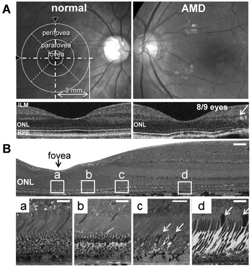
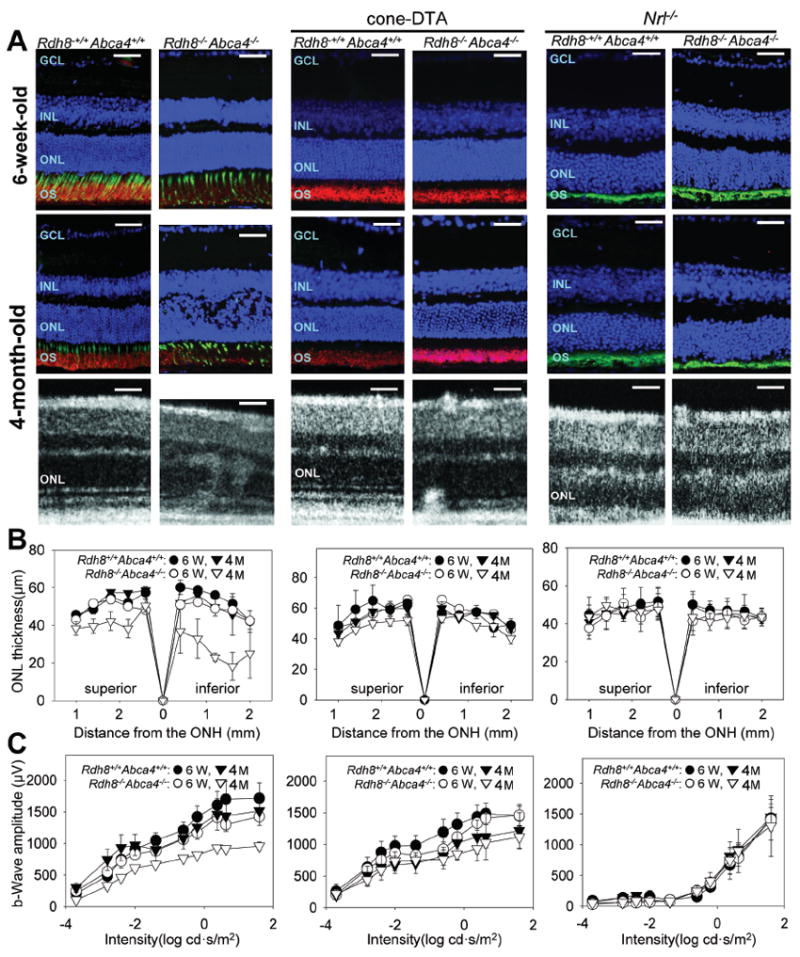
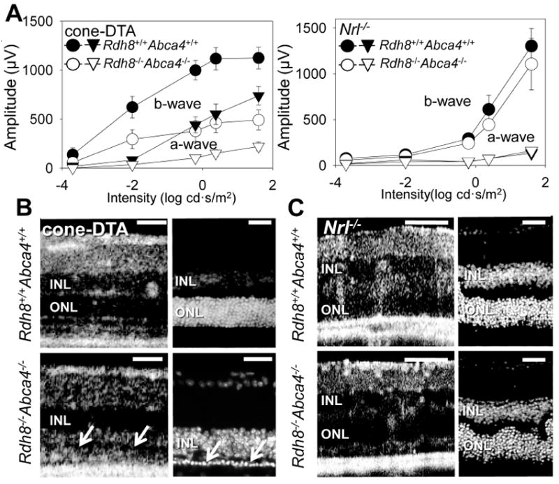
All-trans-retinal and its condensation-products can cause retinal degeneration in a light-dependent manner and contribute to the pathogenesis of human macular diseases such as Stargardt's disease and age-related macular degeneration. Although these toxic retinoid by-products originate from rod and cone photoreceptor cells, the contribution of each cell type to light-induced retinal degeneration is unknown. In this study, the primary objective was to learn whether rods or cones are more susceptible to light-induced, all-trans-retinal-mediated damage. Previously, we reported that mice lacking enzymes that clear all-trans-retinal from the retina, ATP-binding cassette transporter 4 and retinol dehydrogenase 8, manifested light-induced retinal dystrophy. We first examined early-stage age-related macular degeneration patients and found retinal degenerative changes in rod-rich rather than cone-rich regions of the macula. We then evaluated transgenic mice with rod-only and cone-like-only retinas in addition to progenies of such mice inbred with Rdh8(-/-) Abca4(-/-) mice. Of all these strains, Rdh8(-/-) Abca4(-/-) mice with a mixed rod-cone population showed the most severe retinal degeneration under regular cyclic light conditions. Intense light exposure induced acute retinal damage in Rdh8(-/-) Abca4(-/-) and rod-only mice but not cone-like-only mice. These findings suggest that progression of retinal degeneration in Rdh8(-/-) Abca4(-/-) mice is affected by differential vulnerability of rods and cones to light.
© 2012 The Authors. Journal of Neurochemistry © 2012 International Society for Neurochemistry.
Conflict of interest statement
Figures
Similar articles
-
Increased cone sensitivity to ABCA4 deficiency provides insight into macular vision loss in Stargardt's dystrophy.Biochim Biophys Acta. 2012 Jul;1822(7):1169-79. doi: 10.1016/j.bbadis.2011.10.007. Epub 2011 Oct 13. Biochim Biophys Acta. 2012. PMID: 22033104 Free PMC article.
-
Retinol dehydrogenase 8 and ATP-binding cassette transporter 4 modulate dark adaptation of M-cones in mammalian retina.J Physiol. 2015 Nov 15;593(22):4923-41. doi: 10.1113/JP271285. Epub 2015 Oct 18. J Physiol. 2015. PMID: 26350353 Free PMC article.
-
Interruption of the visual cycle in a novel animal model induces progressive vision loss resembling Stargardts Disease.Sci Rep. 2024 Dec 28;14(1):30880. doi: 10.1038/s41598-024-81869-y. Sci Rep. 2024. PMID: 39730605 Free PMC article.
-
Defective cone photoreceptor cytoskeleton, alignment, feedback, and energetics can lead to energy depletion in macular degeneration.Prog Retin Eye Res. 2004 Sep;23(5):495-522. doi: 10.1016/j.preteyeres.2004.04.005. Prog Retin Eye Res. 2004. PMID: 15302348 Review.
-
Is Retinal Metabolic Dysfunction at the Center of the Pathogenesis of Age-related Macular Degeneration?Int J Mol Sci. 2019 Feb 11;20(3):762. doi: 10.3390/ijms20030762. Int J Mol Sci. 2019. PMID: 30754662 Free PMC article. Review.
Cited by
-
The human rhodopsin kinase promoter in an AAV5 vector confers rod- and cone-specific expression in the primate retina.Hum Gene Ther. 2012 Oct;23(10):1101-15. doi: 10.1089/hum.2012.125. Epub 2012 Sep 20. Hum Gene Ther. 2012. PMID: 22845794 Free PMC article.
-
Expression and light-triggered movement of rhodopsins in the larval visual system of mosquitoes.J Exp Biol. 2015 May;218(Pt 9):1386-92. doi: 10.1242/jeb.111526. Epub 2015 Mar 6. J Exp Biol. 2015. PMID: 25750414 Free PMC article.
-
Prospective Cohort Study of Childhood-Onset Stargardt Disease: Fundus Autofluorescence Imaging, Progression, Comparison with Adult-Onset Disease, and Disease Symmetry.Am J Ophthalmol. 2020 Mar;211:159-175. doi: 10.1016/j.ajo.2019.11.008. Epub 2019 Dec 6. Am J Ophthalmol. 2020. PMID: 31812472 Free PMC article.
-
Chemistry and biology of the initial steps in vision: the Friedenwald lecture.Invest Ophthalmol Vis Sci. 2014 Oct 22;55(10):6651-72. doi: 10.1167/iovs.14-15502. Invest Ophthalmol Vis Sci. 2014. PMID: 25338686 Free PMC article.
-
Scavenging of Cation Radicals of the Visual Cycle Retinoids by Lutein, Zeaxanthin, Taurine, and Melanin.Int J Mol Sci. 2023 Dec 29;25(1):506. doi: 10.3390/ijms25010506. Int J Mol Sci. 2023. PMID: 38203675 Free PMC article.
References
-
- Allikmets R. Retinal Degenerations: Biology, Diagnostics and Therapeutics. Humana Press; Totowa, N.J: 2007. Stargardt disease: from gene discovery to therapy; pp. 105–118.
-
- Chen J, Nathans J. Genetic ablation of cone photoreceptors eliminates retinal folds in the retinal degeneration 7 (rd7) mouse. Investigative ophthalmology & visual science. 2007;48:2799–2805. - PubMed
-
- Cideciyan AV, Aleman TS, Swider M, et al. Mutations in ABCA4 result in accumulation of lipofuscin before slowing of the retinoid cycle: a reappraisal of the human disease sequence. Human molecular genetics. 2004;13:525–534. - PubMed
Publication types
MeSH terms
Substances
Grants and funding
LinkOut - more resources
Full Text Sources
Medical
Molecular Biology Databases
