. 2012 Jun;121(5):717-29.
doi: 10.1111/j.1471-4159.2012.07648.x. Epub 2012 Feb 6.

The Cbln family of proteins interact with multiple signaling pathways

Affiliations

The Cbln family of proteins interact with multiple signaling pathways

Peng Wei et al. J Neurochem. 2012 Jun.

Abstract

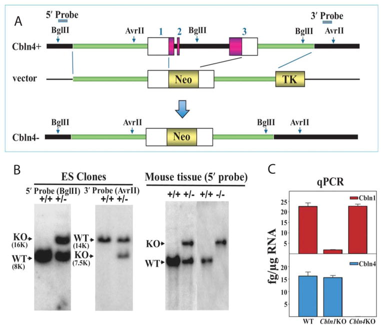
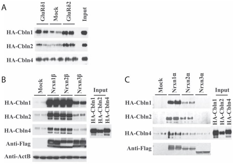
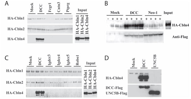
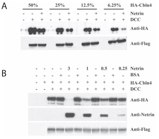
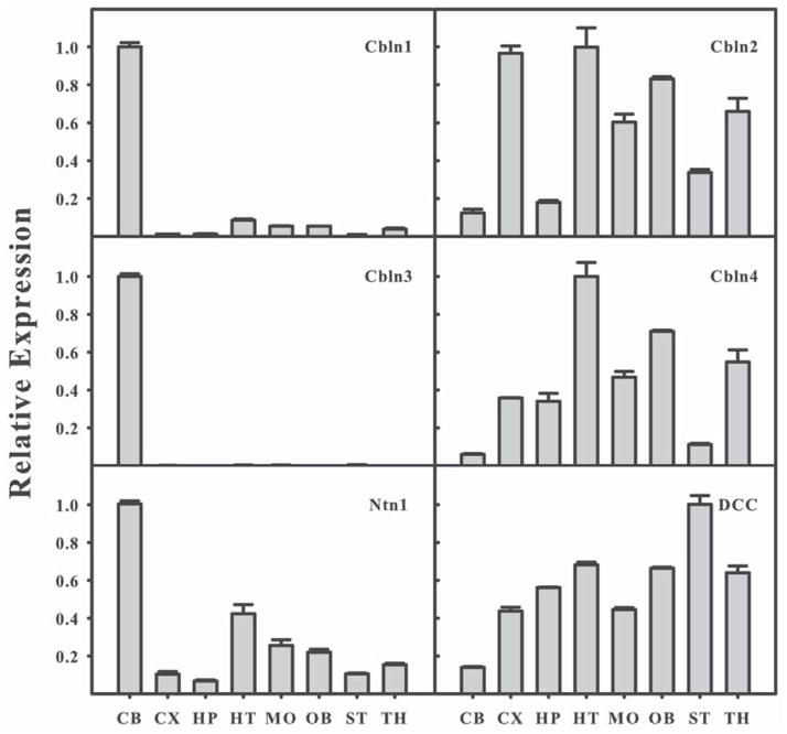
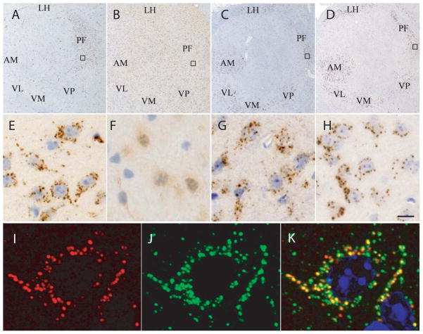
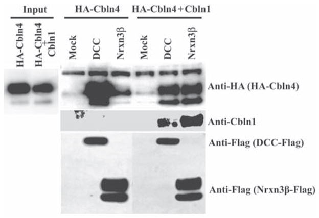
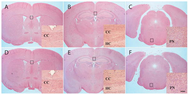
Cerebellin precursor protein (Cbln1) is essential for synapse integrity in cerebellum through assembly into complexes that bridge pre-synaptic β-neurexins (Nrxn) to post-synaptic GluRδ2. However, GluRδ2 is largely cerebellum-specific, yet Cbln1 and its little studied family members, Cbln2 and Cbln4, are expressed throughout brain. Therefore, we investigated whether additional proteins mediate Cbln family actions. Whereas Cbln1 and Cbln2 bound to GluRδ2 and Nrxns1-3, Cbln4 bound weakly or not at all, suggesting it has distinct binding partners. In a candidate receptor-screening assay, Cbln4 (but not Cbln1 or Cbln2) bound selectively to the netrin receptor, (deleted in colorectal cancer (DCC) in a netrin-displaceable fashion. To determine whether Cbln4 had a netrin-like function, Cbln4-null mice were generated. Cbln4-null mice did not phenocopy netrin-null mice. Cbln1 and Cbln4 were likely co-localized in neurons thought to be responsible for synaptic changes in striatum of Cbln1-null mice. Furthermore, complexes containing Cbln1 and Cbln4 had greatly reduced affinity to DCC but increased affinity to Nrxns, suggesting a functional interaction. However, Cbln4-null mice lacked the striatal synaptic changes seen in Cbln null mice. Thus, Cbln family members interact with multiple receptors/signaling pathways in a subunit composition-dependent manner and have independent functions with Cbln4 potentially involved in the less well-characterized role of netrin/DCC in adult brain.


Conflict of interest statement

None of the authors have any financial interests or conflicts of interest in the data presented in the manuscript.

Figures

Fig. 1
Fig. 1
Generation of Cbln4-null mice. (A) Schematic representations of Cbln4 genomic DNA (Cbln4+), targeting vector and targeted gene (Cbln4−). Shaded bars indicate the 5′ and 3′ probes for Southern blot analysis. The region spanning from start codon through to stop codon of Cbln4 was deleted by inserting the neomycin resistance gene cassette (Neo). (B) Southern blot analysis of genomic DNA prepared from ES cells (left and middle panels) and tissues of wild-type (+/+), heterozygote (+/−), and knockout (−/−) mice (right panel). DNA from ES cells and mouse tissues was digested with BglII (for 5′ probe) and AseI (for 3′ probe), analyzed using a digoxigenin (DIG)-labeled 0.5 kb 5′ external probe and a 0.36 kb 3′ probe, respectively. Bands with the indicated sizes denote correct targeting in both ES cells and tissues. (C) qPCR showing loss of Cbln1 (red) and Cbln4 mRNA (blue) in Cbln1-null (Cbln1KO) and Cbln4-null (Cbln4KO) mouse brain, respectively. There is no compensatory change of Cbln1 or Cbln4 mRNA levels in Cbln4- or Cbln1-null mice, respectively.
Fig. 2
Fig. 2
Comparison of binding of HA-tagged Cbln family members to GluRδ1, GluRδ2, and α- and β-neurexins. (A) HEK293T cells were transfected with GluRδ1, GluRδ2 or empty vector (Mock). Cells were subsequently exposed to conditioned medium (Input) containing HA-Cbln1, HA-Cbln2, or HA-Cbln4 for 4 h. After washing with medium, cells were lysed with sample buffer and subjected to SDS-PAGE and immunoblotted with anti-hemagglutinin (HA) antiserum. HA-Cbln1 binds both GluRδ1 and GluRδ2. HA-Cbln2 binds to GluRδ2 but weakly or not at all to GluRδ1. HA-Cbln4 does not bind GluRδ1 or GluRδ2. (B) HEK293T cells were transfected with Nrxn1β, Nrxn2β, Nrxn3β or empty vector (Mock). Then cells were incubated with conditioned medium (Input) containing HA-Cbln1, HA-Cbln2, or HA-Cbln4 for 4 h. After washing with medium, cells were lysed with sample buffer and subjected to SDS-PAGE and immunoblotted with anti-hemagglutinin (HA), anti-Flag or anti-β-actin (ActB) antisera. Compared with mock transfection, both Cbln1 and Cbln2 show binding to Nrxn1β, Nrxn2β and Nrxn3β whereas HA-Cbln4 binds to Nrxn1β and Nrxn2β but not Nrxn3β. (C) HEK293T cells were transfected with Nrxn1α, Nrxn2α, Nrxn3α or empty vector (Mock). Then cells were incubated with conditioned medium (Input) containing HA-Cbln1, HA-Cbln2, or HA-Cbln4 for 4 h. After washing with medium, cells were lysed with sample buffer and subjected to SDS-PAGE and immunoblotted with anti-hemagglutinin (HA) or anti-Flag tag antisera. Compared with mock transfection, both HA-Cbln1 and HA-Cbln2 show binding to Nrxn1α and Nrxn2α, but not Nrxn3α, whereas HA-Cbln4 binds only to Nrxn1α.
Fig. 3
Fig. 3
HA-Cbln4 but not HA-Cbln1 or HA-Cbln2 binds to DCC. (A) HEK293T cells were transfected with DCC, Fcgr1, Cntn3, Ptprg or empty vector (Mock). Then cells were incubated with conditioned medium (Input) containing HA-Cbln1, HA-Cbln2, or HA-Cbln4 for 4 h. After washing with medium, cells were lysed with sample buffer and subjected to SDS-PAGE and immunoblotted with anti-hemagglutinin (HA) antiserum. HA-Cbln4 bound to DCC but not other membrane proteins. Neither HA-Cbln1 nor HA-Cbln2 bound to any of the membrane proteins tested. (B) Neogenin (Neo-1) is not a receptor for Cbln4. HEK-293 cells were transfected with empty vector (Mock), DCC or Neo-1 containing a C-terminal FLAG tag. Cells were subsequently exposed to conditioned medium from mock transfected (−) or HA-Cbln4-transfected (+) cells (Input) for 4 hours, washed with medium, lysed in loading buffer, subjected to SDS-PAGE and immunoblotted with anti-hemagglutinin (HA) or anti-Flag tag antisera. Whereas HA-Cbln4 bound to DCC it did not bind to cells transfected with Neo-1. (C) Cbln family members do not bind to other DCC family members. HEK293T cells were transfected with DCC, Igdcc3, Igdcc4, Igdcc5, Robo1 or empty vector (Mock). Cells were subsequently exposed to conditioned medium (Input) containing HA-Cbln1, HA-Cbln2, or HA-Cbln4 for 4 h. After washing with medium, cells were lysed with sample buffer and subjected to SDS-PAGE and immunoblotted with anti-hemagglutinin (HA) antisera. HA-Cbln4 bound to DCC but no other proteins whereas HA-Cbln1 and HA-Cbln2 did not bind to any of the membrane proteins tested. (D) UNC5B is not a receptor for Cbln4. HEK-293 cells were transfected with empty vector (Mock), DCC or UNC5B and subsequently exposed to conditioned medium containing HA-Cbln4 (Input) for 4 hours, washed with medium, lysed in loading buffer, subjected to SDS-PAGE and immunoblotted with anti-hemagglutinin (HA) or anti-Flag tag antisera. Whereas HA-Cbln4 bound to DCC it did not bind to cells transfected with UNC5B.
Fig. 4
Fig. 4
Netrin-1 competes with HA-Cbln4 for DCC binding. (A) HEK293T cells were transfected with DCC (+) or empty vector (−). Cells were subsequently exposed to varying dilutions (as a %) of conditioned medium containing HA-Cbln4 with (+) or without (−) netrin-1 (1μg/ml). Subsequently cells were harvested and processed for immunoblotting as in Figure 2. The presence of netrin-1 reduced HA-Cbln4 binding to DCC at all dilutions of HA-Cbln4 tested. (B) Cells were transfected with DCC (+) or empty vector (−). Cells were subsequently pre-incubated with varying concentrations of netrin-1 (3, 1, 0.5, 0.25 or 0 μg/ml) or BSA carrier for 30 min, and then incubated for an additional 3.5 hours with conditioned medium containing HA-Cbln4 and the appropriate level of netrin-1 or BSA. Netrin-1 competed with HA-Cbln4 for DCC binding in a dose-dependent manner.
Fig. 5
Fig. 5
Relative expression levels of mRNAs of Cbln1, Cbln2, Cbln3, Cbln4, Ntn1 and DCC in different adult mouse brain regions. Levels of mRNAs were determined by qPCR. The abundance of mRNA was normalized to β-actin and is expressed as the magnitude of the change in abundance relative to the brain region with the highest level for that transcript. CB, cerebellum; CX, cerebral cortex; HP, hippocampus; HT, hypothalamus; MO, medulla oblongata; OB, olfactory bulb; ST, striatum; TH, thalamus. Results expressed as mean ±SE of measurements of 3 samples from independent mice.
Fig. 6
Fig. 6
Localization of Cbln4-like and Cbln1-like immunoreactivities in thalamus. As the anti-Cbln4 antibody used in this experiment cross-reacted with Cbln1 in our hands, the brains from Cbln1-null mice were used in the immunohistochemistry studies to localize the expression of Cbln4. (A) In sagittal sections of adult Cbln1-null moue brains, Cbln4-like immunoreactivity is observed in a punctate pattern (E is enlargement of box in A) in the cytoplasm of thalamic neurons. The most prominent staining is seen in neurons of the parafascicular nucleus of thalamus (PF). (B) Specificity of the anti-Cbln4 antiserum is shown by the lack of Cbln4-like immunoreactivity in thalamic neurons from Cbln1-/Cbln4-double null mice (F is enlargement of box in B). (C) Compared with the localization of Cbln4, Cbln1-like immunoreactivity in a wild type mouse is more widespread in the thalamus with staining observed in not only PF neurons but also neurons in the ventral (VL, VM, VP) and anterior thalamus (AM). Like Cbln4, Cbln1-like immunoreactivity is punctate (G is enlargement of box in C) and of roughly the same incidence as for Cbln4. (D) In Cbln4-null mice there is no overt change in the level or distribution of Cbln1-like immunoreactivity (H is enlargement of box in D). (I–K) Double immunofluorescence labeling was performed on sections from thalamus of Cbln1-null mice using the anti-Cbln4 antiserum (red, I and K) and an antiserum to the lysosome marker cathepsin D (green, J and K). Merged confocal images (K) revealed that the Cbln4 vesicle-like staining almost always co-localizes with cathepsin D (yellow staining). Scale bar: A–D, 360 μm; E–H, 20 μm; I–K, 4 μm. AM, anteromedial thalamic nucleus; LH, lateral habenular nucleus; PF, parafascicular nucleus; VL, ventrolateral thalamic nucleus; VM, ventromedial thalamic nucleus; VP, ventral posterior tier thalamic nuclei.
Fig. 7
Fig. 7
Co-expression of Cbln1 with Cbln4 alters the receptor binding specificity of HA-Cbln4. HEK293T cells were transfected with DCC, Nrxn3β or empty vector (Mock). Cells were subsequently exposed to conditioned medium (Input) derived from HEK293 cells co-transfected with either HA-Cbln4 and mock plasmid or HA-Cbln4 and Cbln1 for 4 h. After washing with medium, cells were lysed with sample buffer and subjected to SDS-PAGE and immunoblotted with anti-hemagglutinin (HA), anti-Cbln1 or anti-Flag tag antisera. HA-Cbln4 shows strong binding to DCC and minimal binding to Nrxn3β. Co-expression of Cbln1 with HA-Cbln4 alters the binding specificity of HA-Cbln4 by reducing its association with DCC and promoting its association with Nrxn3β.
Fig. 8
Fig. 8
Cbln4-null mice have no obvious neuroanatomical abnormalities. Representative matched coronal sections from wild type (A–C) or Cbln4-null (D–E) mice were stained with hematoxylin and eosin. The sections were selected to highlight regions of brain (boxes in A–F) that have anomalies in netrin- and DCC-null mice. Compared with wild-type (A–C) mice, Cbln4-null mice (D–F) show no overt neuroanatomical defects. Furthermore, specific structures such as corpus callosum (CC in D, E), hippocampal commissure (HC, in E), and pontine nuclei (PN in F) that are lacking in netrin- and DCC-null mice are present and indistinguishable from wild type littermates in Cbln4-null mice. Scale bar, 750 μm for A–F and 100 μm for the insets.
Fig. 9
Fig. 9
Cbln4-null mice do not exhibit the same deficits as Cbln1-null mice. (A) Cbln4-null mice (open circles, KO) are indistinguishable from wild type (filled circles, WT) gender-matched littermates (p>0.5, n=9/genotype) on the accelerating rotarod assay. Data is mean ± SEM. (B) There is no difference in dendritic spine densities on striatal medium spiny neurons of Cbln4-null (KO) compared to wild type (WT) littermates (p>0.1, n=4/genotype).

Comment in

Similar articles

Cited by

References

    1. Ackerman SL, Kozak LP, Przyborski SA, Rund LA, Boyer BB, Knowles BB. The mouse rostral cerebellar malformation gene encodes an UNC-5-like protein. Nature. 1997;386:838–842. - PubMed
    1. Andrews GL, Tanglao S, Farmer WT, Morin S, Brotman S, Berberoglu MA, Price H, Fernandez GC, Mastick GS, Charron F, Kidd T. Dscam guides embryonic axons by Netrin-dependent and independent functions. Development. 2008;135:3839–3848. - PMC - PubMed
    1. Araki K, Meguro H, Kushiya E, Takayama C, Inoue Y, Mishina M. Selective expression of the glutamate receptor channel delta 2 subunit in cerebellar Purkinje cells. Biochem Biophys Res Commun. 1993;197:1267–1276. - PubMed
    1. Bao D, Pang Z, Morgan MA, Parris J, Rong Y, Li L, Morgan JI. Cbln1 is essential for interaction-dependent secretion of Cbln3. Mol Cell Biol. 2006;26:9327–9337. - PMC - PubMed
    1. Bao D, Pang Z, Morgan JI. The structure and proteolytic processing of Cbln1 complexes. J Neurochem. 2005;95:618–629. - PubMed

Publication types