

Palmitic and linoleic acids induce ER stress and apoptosis in hepatoma cells
- PMID: 22221411
- PMCID: PMC3306830
- DOI: 10.1186/1476-511X-11-1
Palmitic and linoleic acids induce ER stress and apoptosis in hepatoma cells
Abstract
Objectives: Hepatic inflammation and degeneration induced by lipid depositions may be the major cause of nonalcoholic fatty liver disease. In this study, we tried to investigate the effects of saturated and unsaturated fatty acids on hepatoma cell apoptosis.
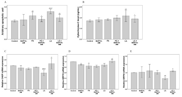
Methods: H4IIE liver cells were treated with palmitic acid, linoleic acid, or both with or without the calcium-specific chelator BAPTA-AM after which the expression of proteins associated with endoplasmic reticulum (ER) stress, apoptosis, caspase-3 levels, and calcium flux were measured.
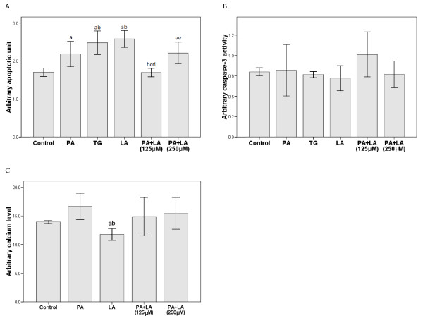
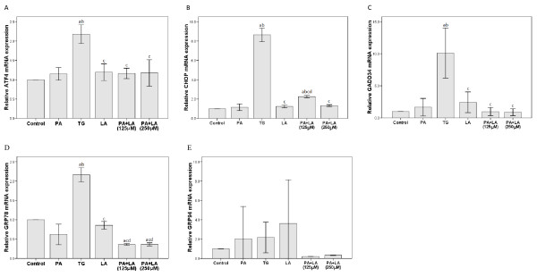
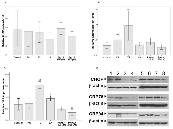
Results: Palmitic or linoleic acid (250 μM) induced H4IIE cell apoptosis, which required calcium flux but not caspase-3. Apoptosis was not observed when cells were co-treated with linoleic acid (125 μM) and palmitic acid (250 μM). Importantly, the release of cytochrome C from mitochondria into cytoplasm during cell apoptosis was specifically detected only when linoleic acid (125 μM), but not palmitic acid (250 μM), was added to the cells. Depletion of intracellular calcium flux by the calcium-specific chelator, BAPTA-AM, abolished linoleic acid-induced apoptosis. Moreover, in the presence of BAPTA-AM, expression of the unfolded protein response (UPR)-associated genes, CHOP, GRP78, and GRP94, was induced by linoleic acid, but not palmitic acid.
Conclusions: The results suggest that linoleic acid promotes cell apoptosis through the release of cytochrome C, only if the intracellular calcium flux is unperturbed and intact. These results confirm that ER stress contributes to fatty acid-induced liver cell apoptosis.
Figures
Similar articles
-
Effect of α-linolenic acid on endoplasmic reticulum stress-mediated apoptosis of palmitic acid lipotoxicity in primary rat hepatocytes.Lipids Health Dis. 2011 Jul 25;10:122. doi: 10.1186/1476-511X-10-122. Lipids Health Dis. 2011. PMID: 21787405 Free PMC article.
-
Polychlorinated biphenyl quinone induces endoplasmic reticulum stress, unfolded protein response, and calcium release.Chem Res Toxicol. 2015 Jun 15;28(6):1326-37. doi: 10.1021/acs.chemrestox.5b00124. Epub 2015 May 18. Chem Res Toxicol. 2015. PMID: 25950987
-
Ethanol promotes saturated fatty acid-induced hepatoxicity through endoplasmic reticulum (ER) stress response.Chin J Nat Med. 2015 Apr;13(4):250-6. doi: 10.1016/S1875-5364(15)30011-X. Chin J Nat Med. 2015. PMID: 25908621
-
Fatty acids and the endoplasmic reticulum in nonalcoholic fatty liver disease.Biofactors. 2011 Jan-Feb;37(1):8-16. doi: 10.1002/biof.135. Epub 2010 Dec 2. Biofactors. 2011. PMID: 21328622 Free PMC article. Review.
-
Induction of Hepatoma Cell Pyroptosis by Endogenous Lipid Geranylgeranoic Acid-A Comparison with Palmitic Acid and Retinoic Acid.Cells. 2024 May 9;13(10):809. doi: 10.3390/cells13100809. Cells. 2024. PMID: 38786033 Free PMC article. Review.
Cited by
-
Linoleic Acid-Induced Growth Inhibition of Human Gastric Epithelial Adenocarcinoma AGS Cells is Associated with Down-Regulation of Prostaglandin E2 Synthesis and Telomerase Activity.J Cancer Prev. 2014 Mar;19(1):31-8. doi: 10.15430/jcp.2014.19.1.31. J Cancer Prev. 2014. PMID: 25337570 Free PMC article.
-
Calcium signaling in the liver.Compr Physiol. 2013 Jan;3(1):515-39. doi: 10.1002/cphy.c120013. Compr Physiol. 2013. PMID: 23720295 Free PMC article. Review.
-
LRRK2 Regulates CPT1A to Promote β-Oxidation in HepG2 Cells.Molecules. 2020 Sep 9;25(18):4122. doi: 10.3390/molecules25184122. Molecules. 2020. PMID: 32916992 Free PMC article.
-
Induction of fat apoptosis by a non-thermal device: Mechanism of action of non-invasive high-intensity electromagnetic technology in a porcine model.Lasers Surg Med. 2019 Jan;51(1):47-53. doi: 10.1002/lsm.23039. Epub 2018 Dec 14. Lasers Surg Med. 2019. PMID: 30549290 Free PMC article.
-
Anti-Rheumatoid Arthritis Pharmacodynamic Substances Screening of Periploca forrestii Schltr.: Component Analyses In Vitro and In Vivo Combined with Multi-Technical Metabolomics.Int J Mol Sci. 2023 Sep 5;24(18):13695. doi: 10.3390/ijms241813695. Int J Mol Sci. 2023. PMID: 37761998 Free PMC article.
References
-
- Solís Herruzo JA, García Ruiz I, Pérez Carreras M, Muñoz Yagüe MT. Non-alcoholic fatty liver disease. From insulin resistance to mitochondrial dysfunction. Rev Esp Enferm Dig. 2006;98:844–874. - PubMed
-
- Artwohl M, Lindenmair A, Roden M, Waldhäusl WK, Freudenthaler A, Klosner G, Ilhan A. et al.Fatty acids induce apoptosis in human smooth muscle cells depending on chain length, saturation, and duration of exposure. Atherosclerosis. 2009;202:351–362. doi: 10.1016/j.atherosclerosis.2008.05.030. - DOI - PubMed
MeSH terms
Substances
LinkOut - more resources
Full Text Sources
Other Literature Sources
Research Materials
Miscellaneous
