

fMRI responses to emotional faces in children and adolescents at genetic risk for psychiatric illness share some of the features of depression
- PMID: 22222174
- PMCID: PMC5166711
- DOI: 10.1016/j.jad.2011.11.036
fMRI responses to emotional faces in children and adolescents at genetic risk for psychiatric illness share some of the features of depression
Abstract
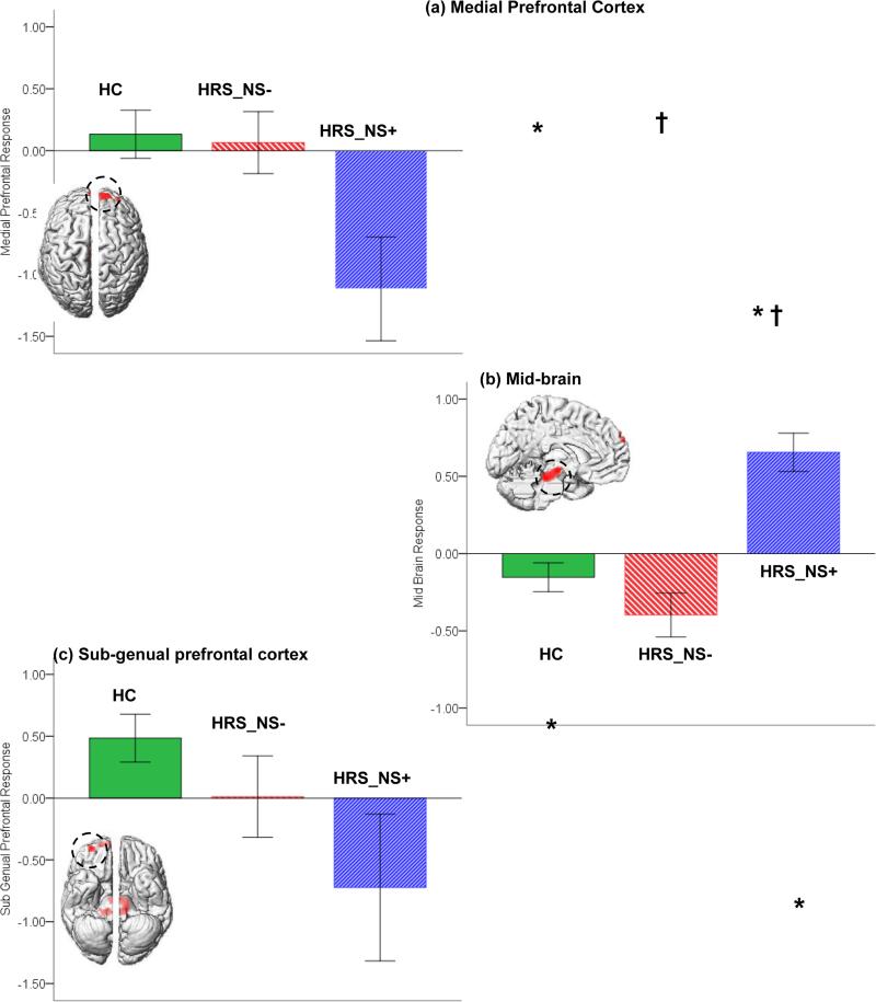
Background: Fronto-limbic regions of the brain including the sub-genual (sgPFC) and medial prefrontal (mPFC) cortices are central to processing emotionally salient and hedonic stimuli (Mayberg, 2009) and implicated in depression. The relevance of cortico-limbic models of emotion and reward processing in children with genetic risk for psychiatric disorders has not been assessed.
Methods: Here we studied adolescents at risk for schizophrenia (HRS) and controls (HC) using an event-related fMRI continuous affective appraisal task. HRS were divided into sub-groups based on the presence or absence of negative symptoms (Miller et al., 2003), HRS_NS+ and HRS_NS- respectively. Brain responses to positive, negative and neutral emotional stimuli were estimated.
Results: Consistent with observations in the depressive phenotype, for positively valenced stimuli, HRS_NS+ (relative to HC and HRS_NS-) were characterized by hypo-responsivity of the sgPFC and the mPFC, but hyper-responsivity of the mid-brain. sgPFC and mPFC signals were coupled across groups.
Limitations: Such studies can benefit from larger sample sizes, though our observed effect sizes were in the moderate to large range.
Conclusions: Children and adolescents at risk for psychiatric illness and who evince reliably present negative symptoms show brain responses to socially rewarding stimuli similar to those observed in depression. Studies in at-risk children and adolescents may be important in understanding how early manifestations of depression-like characteristics impact brain function.
Copyright © 2011 Elsevier B.V. All rights reserved.
Figures


Similar articles
-
Altered amygdala-prefrontal connectivity during emotion perception in schizophrenia.Schizophr Res. 2016 Aug;175(1-3):35-41. doi: 10.1016/j.schres.2016.04.003. Epub 2016 Apr 12. Schizophr Res. 2016. PMID: 27083779
-
Lack of cortico-limbic coupling in bipolar disorder and schizophrenia during emotion regulation.Transl Psychiatry. 2012 Mar 13;2(3):e90. doi: 10.1038/tp.2012.16. Transl Psychiatry. 2012. PMID: 22832855 Free PMC article.
-
Altered medial prefrontal activity during dynamic face processing in schizophrenia spectrum patients.Schizophr Res. 2014 Aug;157(1-3):225-30. doi: 10.1016/j.schres.2014.05.023. Epub 2014 Jun 2. Schizophr Res. 2014. PMID: 24888525
-
Functional atlas of emotional faces processing: a voxel-based meta-analysis of 105 functional magnetic resonance imaging studies.J Psychiatry Neurosci. 2009 Nov;34(6):418-32. J Psychiatry Neurosci. 2009. PMID: 19949718 Free PMC article. Review.
-
Neurocognitive correlates of emotional stimulus processing in pediatric bipolar disorder: a review.Postgrad Med. 2010 Jul;122(4):94-104. doi: 10.3810/pgm.2010.07.2177. Postgrad Med. 2010. PMID: 20675973 Review.
Cited by
-
Lower prefrontal activation during emotion regulation in subjects at ultrahigh risk for psychosis: an fMRI-study.NPJ Schizophr. 2015 Sep 23;1:15026. doi: 10.1038/npjschz.2015.26. eCollection 2015. NPJ Schizophr. 2015. PMID: 27336036 Free PMC article.
-
Cognitive Brain Signatures of Youth With Early Onset and Relatives With Schizophrenia: Evidence From fMRI Meta-analyses.Schizophr Bull. 2020 Jul 8;46(4):857-868. doi: 10.1093/schbul/sbz130. Schizophr Bull. 2020. PMID: 31978222 Free PMC article.
-
Neural antecedents of emotional disorders: a functional magnetic resonance imaging study of subsyndromal emotional symptoms in adolescent girls.Biol Psychiatry. 2013 Aug 15;74(4):265-72. doi: 10.1016/j.biopsych.2013.01.030. Epub 2013 Mar 7. Biol Psychiatry. 2013. PMID: 23477296 Free PMC article.
-
Epigenetics, stress, and their potential impact on brain network function: a focus on the schizophrenia diatheses.Front Psychiatry. 2014 Jun 23;5:71. doi: 10.3389/fpsyt.2014.00071. eCollection 2014. Front Psychiatry. 2014. PMID: 25002852 Free PMC article. Review.
-
Impact of cross-disorder polygenic risk on frontal brain activation with specific effect of schizophrenia risk.Schizophr Res. 2015 Feb;161(2-3):484-9. doi: 10.1016/j.schres.2014.10.046. Epub 2014 Nov 20. Schizophr Res. 2015. PMID: 25468172 Free PMC article.
References
-
- Amodio DM, Frith CD. Meeting of minds: the medial frontal cortex and social cognition. Nat. Rev. Neurosci. 2006;7:268–277. - PubMed
-
- Anand A, Li Y, Wang Y, Wu J, Gao S, Bukhari L, Mathews VP, Kalnin A, Lowe MJ. Activity and connectivity of brain mood regulating circuit in depression: a functional magnetic resonance study. Biol. Psychiatry. 2005;57:1079–1088. - PubMed
-
- Bottlender R, Sato T, Groll C, Jager M, Kunze I, Moller HJ. Negative symptoms in depressed and schizophrenic patients: how do they differ? The J. Clin. Psychiatry. 2003;64:954–958. - PubMed
Publication types
MeSH terms
Grants and funding
LinkOut - more resources
Full Text Sources
Medical
Miscellaneous
