

Postsynaptic signaling during plasticity of dendritic spines
- PMID: 22222350
- PMCID: PMC3306839
- DOI: 10.1016/j.tins.2011.12.002
Postsynaptic signaling during plasticity of dendritic spines
Abstract
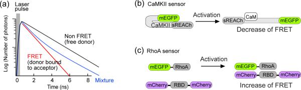
Dendritic spines, small bulbous postsynaptic compartments emanating from neuronal dendrites, have been thought to serve as basic units of memory storage. Despite their small size (~0.1 femtoliter), thousands of species of proteins exist in the spine, including receptors, channels, scaffolding proteins and signaling enzymes. Biochemical signaling mediated by these molecules leads to morphological and functional plasticity of dendritic spines, and ultimately learning and memory in the brain. Here, we review new insights into the mechanisms underlying spine plasticity brought about by recent advances in imaging techniques to monitor molecular events in single dendritic spines. The activity of each protein displays a specific spatiotemporal pattern, coordinating downstream events at different microdomains to change the function and morphology of dendritic spines.
Copyright © 2012 Elsevier Ltd. All rights reserved.
Figures


References
-
- Svoboda K, et al. Direct measurement of coupling between dendritic spines and shafts. Science. 1996;272:716–719. - PubMed
-
- Bloodgood BL, Sabatini BL. Neuronal Activity Regulates Diffusion Across the Neck of Dendritic Spines. Science. 2005;310:866–869. - PubMed
-
- Sabatini BL, et al. The life cycle of Ca2+ ions in dendritic spines. Neuron. 2002;33:439–452. - PubMed
Publication types
MeSH terms
Grants and funding
LinkOut - more resources
Full Text Sources
