Comparative Study
. 2012 Feb;116(2):372-84.
doi: 10.1097/ALN.0b013e318242b2cd.

Ketamine-induced neuroapoptosis in the fetal and neonatal rhesus macaque brain

Affiliations
Comparative Study

Ketamine-induced neuroapoptosis in the fetal and neonatal rhesus macaque brain

Ansgar M Brambrink et al. Anesthesiology. 2012 Feb.

Abstract

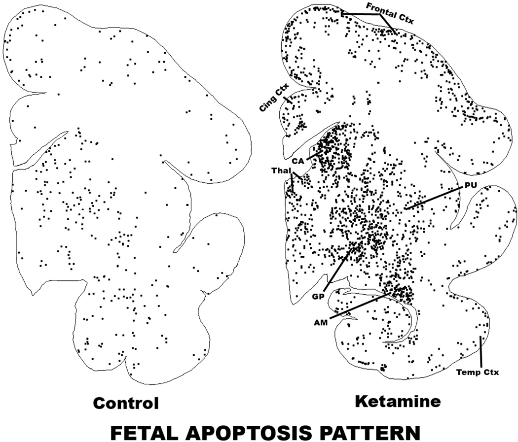
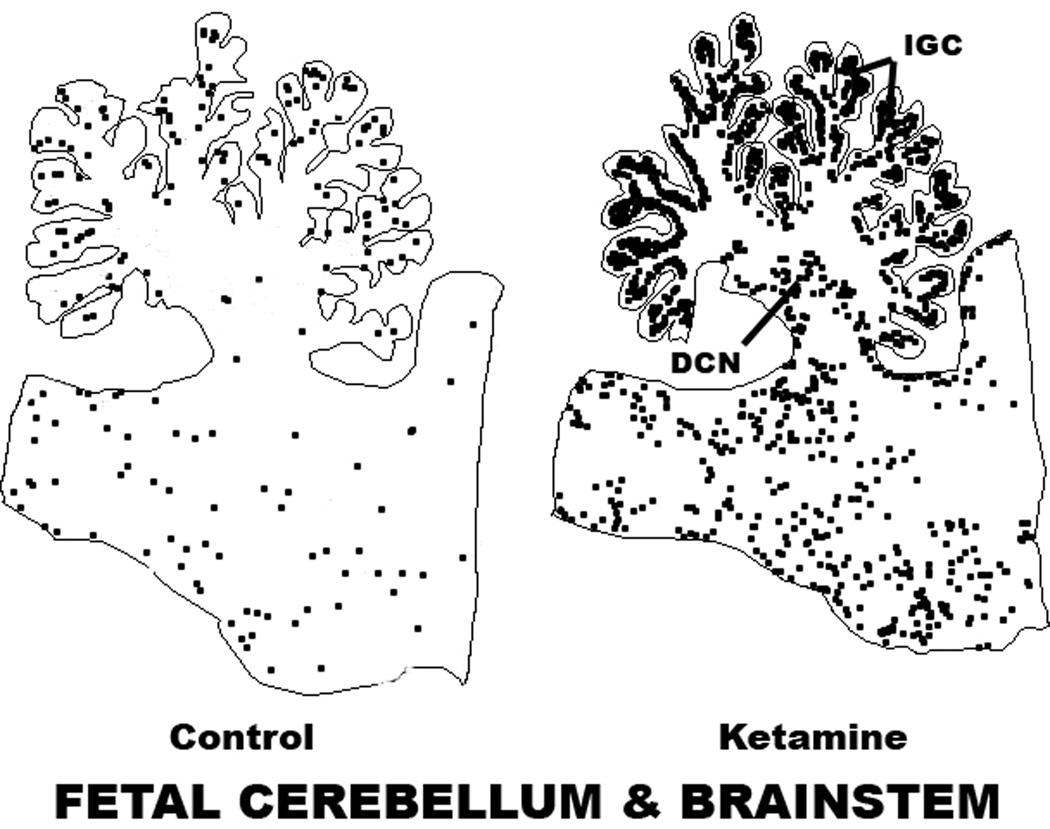
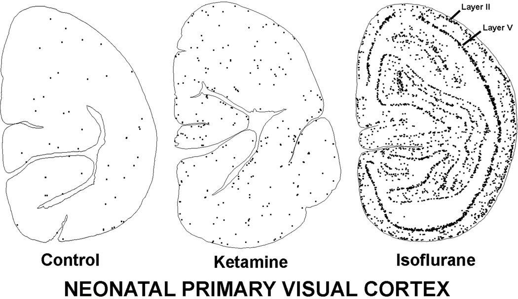
Background: Exposure of rhesus macaque fetuses for 24 h or neonates for 9 h to ketamine anesthesia causes neuroapoptosis in the developing brain. The current study clarifies the minimum exposure required for and the extent and spatial distribution of ketamine-induced neuroapoptosis in rhesus fetuses and neonates.

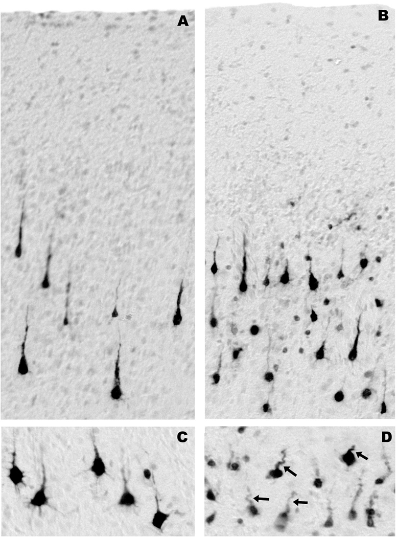
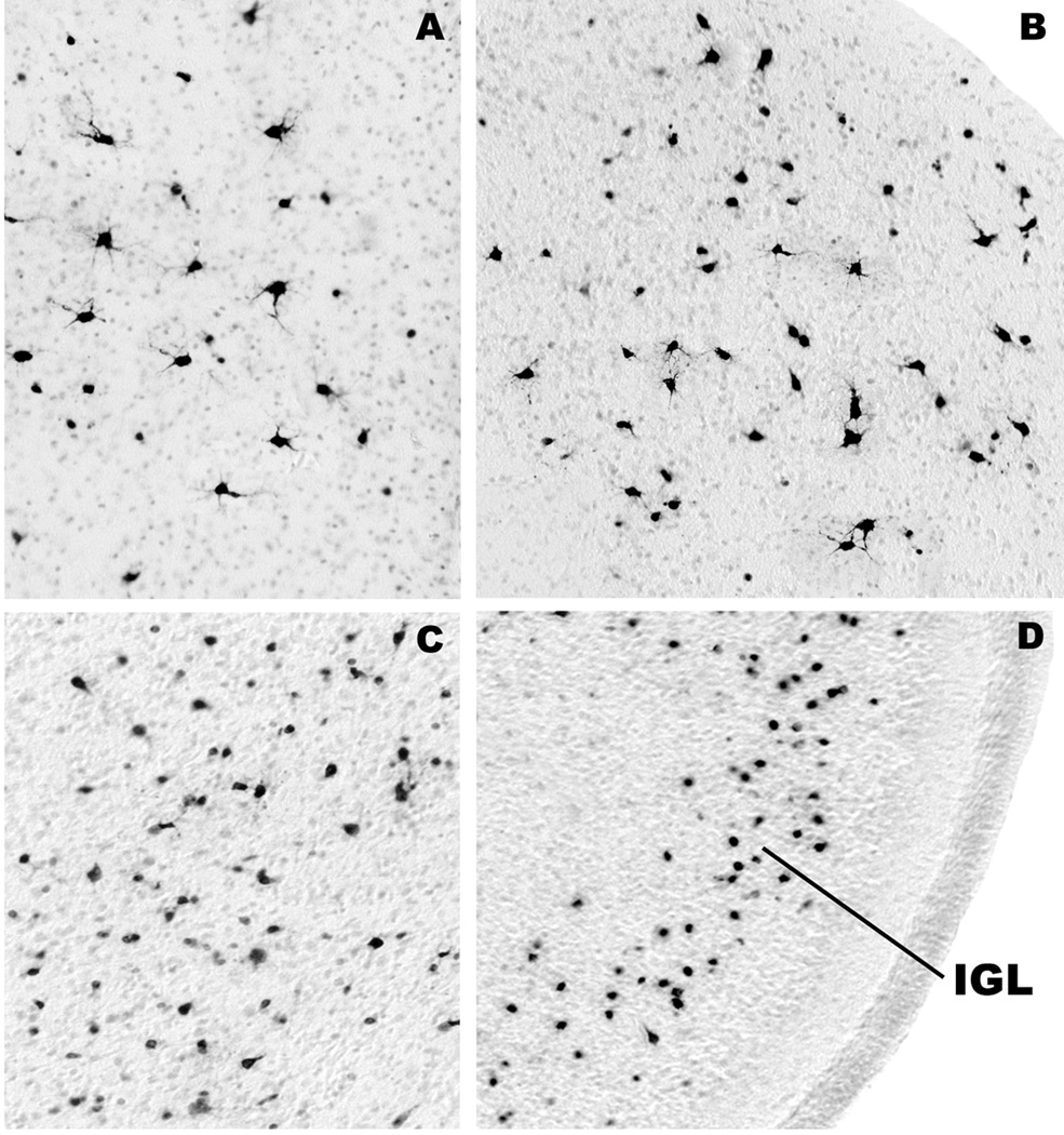
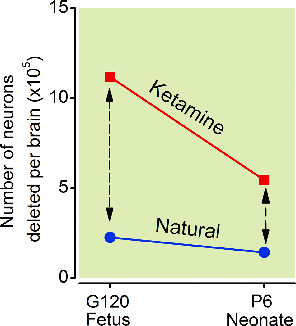
Method: Ketamine was administered by IV infusion for 5 h to postnatal day 6 rhesus neonates or to pregnant rhesus females at 120 days' gestation (full term = 165 days). Three hours later, fetuses were delivered by cesarean section, and the fetal and neonatal brains were studied for evidence of apoptotic neurodegeneration, as determined by activated caspase-3 staining.

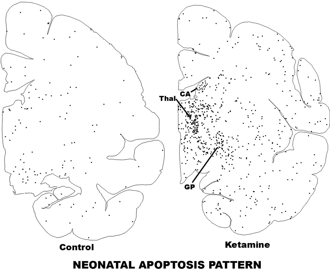
Results: Both the fetal (n = 3) and neonatal (n = 4) ketamine-exposed brains had a significant increase in apoptotic profiles compared with drug-naive controls (fetal n = 4; neonatal n = 5). Loss of neurons attributable to ketamine exposure was 2.2 times greater in fetuses than in neonates. The pattern of neurodegeneration in fetuses was different from that in neonates, and all subjects exposed at either age had a pattern characteristic for that age.

Conclusion: The developing rhesus macaque brain is sensitive to the apoptogenic action of ketamine at both a fetal and neonatal age, and exposure duration of 5 h is sufficient to induce a significant neuroapoptosis response at either age. The pattern of neurodegeneration induced by ketamine in fetuses was different from that in neonates, and loss of neurons attributable to ketamine exposure was 2.2 times greater in the fetal than neonatal brains.


Figures

Figure 1
Figure 1
Neuroapoptosis, as detected by activated caspase-3 (AC3) positivity, induced in the fetal and neonatal monkey brain by ketamine exposure for 5 h. The mean number of AC3-positive neuronal profiles in the ketamine-exposed fetal brains was 4.9-fold higher than in the drug-naive fetal control brains. The difference between the means for ketamine-exposed versus control brains was 8.92 ×10⁵ (95% confidence interval, 13.18 ×10⁵ to 4.65 ×10⁵, p = 0.003). The mean number of AC3-positive neuronal profiles in the ketamine-exposed neonatal brains was 3.83-fold higher than in the drug-naive neonatal control brains. The difference in the mean number of apoptotic neurons between the ketamine and control groups was 4.02 ×10⁵ (95% confidence interval, 6.29 ×10⁵ to 1.74 ×10⁵, p=0.004). AC3 = activated caspase-3.
Figure 2
Figure 2
Computer plot depicting the pattern of neuroapoptosis in ketamine-exposed versus control fetal macaque brains. These sections display several gray matter zones, including frontal, cingulate (Cing) and temporal (Temp) cortices, thalamus (Thal), caudate nucleus (CA), putamen (PU), globus pallidus (GP) and amygdala (AM). An outline of each section was sketched into the computer and the location of each activated caspase-3-stained neuron was marked by a black dot. The pattern of neuronal staining in the control brain due to natural neuroapoptosis is similar to the pattern in the ketamine-exposed brain, but stained neuronal profiles in the former are sparse and in the latter are abundant and heavily concentrated in regions such as the frontal cortex, thalamus, caudate nucleus, globus pallidus and amygdala.
Figure 3
Figure 3
Computer plot showing the pattern of neuroapoptosis in sagittal sections of the cerebellum and brain stem of ketamine-exposed versus control fetal macaque brains. The apoptotic profiles in the cerebellar folia are in the internal granule cell (IGC) zone and those at the base of the cerebellum are in the region of the deep cerebellar nuclei (DCN).
Figure 4
Figure 4
All panels are from fetal brains harvested 3 h after 5 h exposure to ketamine. They display the histological appearance of activated caspase-3 (AC3)-positive neuronal profiles in layers III (A) and V (C) of the frontal cortex, layer III of the temporal cortex (B), and the pyramidal cell layer in the subiculum (D). These panels depict neurons in several stages of degeneration. Subicular neurons are in a late stage of degeneration because they are very sensitive and tend to degenerate early; this is revealed by faint and incomplete staining and by the corkscrew deformity of their apical dendrites (arrows, D). Affected neurons in layer III of the temporal cortex are heterogeneous; some degenerate early and some late. The early dying cells have degenerated beyond recognition and present as faintly stained small round dots (B). Neurons in layer V of the frontal cortex (C) are relatively resistant and have just begun to degenerate. Therefore, they are robustly AC3-positive and show very few signs of structural deformity.
Figure 5
Figure 5
The histological appearance of activated caspase-3-positive neuronal profiles in the lateral septum (A), anterodorsal thalamic nucleus (B), caudate nucleus (C) and cerebellum (D) of fetal brains harvested 3 hours after 5 hours exposure to ketamine. Most of the affected neurons in the septum (A) and thalmus (B) are large multipolar neurons that are relatively resistant and, therefore, are in a relatively early stage of degeneration. The affected neurons in the caudate nucleus (C) are morphologically heterogeneous. A small cell type degenerates very rapidly and presents as a faintly stained, small, condensed, shrunken structure amidst other larger cells that degenerate on a slightly later time schedule. The most sensitive cell population in the cerebellum (D) is one that is located in the inner portion of the inner granule cell layer (IGL). These cells degenerate rapidly following exposure of either infant rodents or fetal monkeys to alcohol or anesthetic drugs. As is explained in previous publications,, it is not clear whether they are granule cells or other cell types that are migrating through the granule cell layer.
Figure 6
Figure 6
Computer plot revealing the pattern of neuroapoptosis in ketamine-exposed versus control neonatal macaque brains. These sections are cut through the forebrain at the same level as the fetal sections in figure 3. Although, the density of apoptotic neural profiles is decreased in the neonatal compared to the fetal brain, the pattern at both ages features a high density of apoptotic profiles in the basal ganglia, including the caudate nucleus (CA), globus pallidus (GP), and adjacent thalamus (Thal) relative to other regions of the neonatal brain.
Figure 7
Figure 7
Apoptosis induced by ketamine vs. isoflurane in the neonatal primary visual cortex. The ketamine illustration is from the present study and the isoflurane illustration is reproduced from our prior publication pertaining to isoflurane-induced neuroapoptosis. The control brain has a very sparse and randomly scattered pattern of neuroapoptosis. The ketamine-exposed visual cortex has a moderately increased number of neural apoptotic profiles and they are distributed in a less random pattern. The isoflurane-exposed visual cortex is heavily studded with apoptotic neural profiles and they are organized in a distinctive laminar pattern corresponding to the location of neurons in layers II and V.
Figure 8
Figure 8
The impact of ketamine treatment on neurons in the fetal versus neonatal brain is depicted in terms of the mean number of neurons affected in the brain by ketamine at each age. For comparison the mean number undergoing natural neuroapoptosis in the control brains is also illustrated. The rate of neuroapoptosis induced by ketamine declines more sharply between the fetal and neonatal period than the rate for natural neuroapoptosis. The dashed lines with arrows represent the amount of apoptosis that can be attributed to ketamine treatment at each age. The number of neurons affected by ketamine exposure in the fetal period is 2.2 times greater than in the neonatal period.

References

    1. Ikonomidou C, Bosch F, Miksa M, Bittigau P, Vöckler J, Dikranian K, Tenkova T, Stevoska V, Turski L, Olney JW. Blockade of NMDA receptors and apoptotic neurodegeneration in the developing brain. Science. 1999;283:70–74. - PubMed
    1. Ikonomidou C, Bittigau P, Ishimaru MJ, Wozniak DF, Koch C, Genz K, Price MT, Stefovska V, Hörster F, Tenkova T, Dikranian K, Olney JW. Ethanol-induced apoptotic neurodegeneration and fetal alcohol syndrome. Science. 2000;287:1056–1060. - PubMed
    1. Olney JW, Tenkova T, Dikranian K, Qin YQ, Labruyere J, Ikonomidou C. Ethanol-induced apoptotic neurodegeneration in the developing C57BL/6 mouse brain. Dev Brain Res. 2002;133:115–126. - PubMed
    1. Bittigau P, Sifringer M, Genz K, Reith E, Pospischil D, Govindarajalu S, Dzietko M, Pesditschek S, Mai I, Dikranian K, Olney JW, Ikonomidou C. Antiepileptic drugs and apoptotic neurodegeneration in the developing brain. Proc Natl Acad Sci USA. 2002;99:15089–15094. - PMC - PubMed
    1. Jevtovic-Todorovic V, Hartman RE, Izumi Y, Benshoff ND, Dikranian K, Zorumski CF, Olney JW, Wozniak DF. Early exposure to common anesthetic agents causes widespread neurodegeneration in the developing rat brain and persistent learning deficits. J Neurosci. 2003;23:876–882. - PMC - PubMed

Publication types