

Quantifying cardiac functions in embryonic and adult zebrafish
- PMID: 22222517
- PMCID: PMC3762588
- DOI: 10.1007/978-1-61779-523-7_2
Quantifying cardiac functions in embryonic and adult zebrafish
Abstract
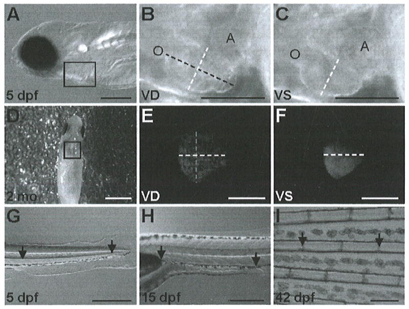
Zebrafish embryos have been extensively used to study heart development and cardiac function, mainly due to the unique embryology and genetics of this model organism. Since most human heart disease occurs during adulthood, adult zebrafish models of heart disease are being created to dissect mechanisms of the disease and discover novel therapies. However, due to its small heart size, the use of cardiac functional assays in the adult zebrafish has been limited. To address this bottleneck, the transparent fish line casper;Tg(cmlc2:nuDsRed) that has a red fluorescent heart can be used to document beating hearts in vivo and to quantify cardiac functions in adult zebrafish. Here, we describe our methods for quantifying shortening fraction and heart rate in embryonic zebrafish, as well as in the juvenile and adult casper;Tg(cmlc2:nuDsRed) fish. In addition, we describe the red blood cell flow rate assay that can be used to reflect cardiac function indirectly in zebrafish at any stage.
Figures
References
-
- Chico TJ, Ingham PW, Crossman DC. Modeling cardiovascular disease in the zebrafish. Trends Cardiovasc Med. 2008;4:150–155. - PubMed
-
- Lieschke GJ, Currie PD. Animal models of human disease: zebrafish swim into view. Nat Rev Genet. 2007;8:353–367. - PubMed
-
- Glickman NS, Yelon D. Cardiac development in zebrafish: coordination of form and function. Semin Cell Dev Biol. 2002;13:507–513. - PubMed
-
- Nasevicius A, Ekker SC. Effective targeted gene ‘knockdown’ in zebrafish. Nat Genet. 2000;26:216–220. - PubMed
MeSH terms
Grants and funding
LinkOut - more resources
Full Text Sources
Miscellaneous
