. 2012 Apr;302(7):G748-57.
doi: 10.1152/ajpgi.00409.2011. Epub 2012 Jan 5.

Hypoxia sensitization of hepatocytes to neutrophil elastase-mediated cell death depends on MAPKs and HIF-1α

Affiliations

Hypoxia sensitization of hepatocytes to neutrophil elastase-mediated cell death depends on MAPKs and HIF-1α

Erica M Sparkenbaugh et al. Am J Physiol Gastrointest Liver Physiol. 2012 Apr.

Abstract

The liver is sensitive to pathological conditions associated with tissue hypoxia (Hx) and the presence of activated neutrophils that secrete the serine protease elastase (EL). We demonstrated previously that cotreatment of rat hepatocytes with nontoxic levels of Hx and EL caused synergistic cell death. Hx is sensed by hypoxia-inducible factor (HIF)-1α, a transcription factor that heterodimerizes with HIF-1β/aryl hydrocarbon receptor nuclear translocator and directs expression of many genes, including the pro-cell death gene Bcl-2/adenovirus E1B-interacting protein 3 (BNIP3). Since cell death from EL or Hx also requires MAPK activation, we tested the hypothesis that the cytotoxic interaction of Hx and EL depends on MAPK and HIF-1α signaling. Treatment of Hepa1c1c7 cells with EL in the presence of Hx (2% O(2)) resulted in synergistic cell death. EL reduced phosphorylated ERK in O(2)-replete and Hx-exposed cells, and ERK inhibition enhanced the cytotoxicity of EL alone. Hx-EL cotreatment caused an additive increase in phosphorylated p38, and p38 inhibition attenuated cell death caused by this cotreatment. EL enhanced Hx-induced HIF-1α accumulation and transcription of the HIF-1α-mediated cell death gene BNIP3, and p38 inhibition attenuated BNIP3 expression and production. Cytotoxicity and BNIP3 expression from EL-Hx cotreatment were reduced in HIF-1β-deficient HepaC4 cells compared with Hepa1c1c7 cells. These results suggest that p38 signaling contributes to Hx-EL cotreatment-induced cell death via modulation of HIF-1α-mediated gene transcription. Finally, lipid peroxidation was enhanced in Hx-EL-cotreated cells compared with cells treated with EL or Hx alone. Vitamin E treatment attenuated lipid peroxidation and protected cells from the cytotoxicity of Hx and EL, suggesting that lipid peroxidation plays a role.


Figures

Fig. 1.
Fig. 1.
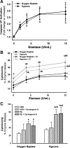
Hypoxia sensitizes primary hepatic parenchymal cells (HPCs) and Hepa1c1c7 cells to the cytotoxicity of elastase (EL). A: primary rat HPCs were treated with 0, 8.5, or 17 U/ml EL in an O2-replete [OxR (20% O2 and 5% CO2, 37°C)] or a hypoxic [Hx (5% O2 in the atmosphere)] environment for 8 h, and cytotoxicity was assessed from release of alanine aminotransferase (ALT) into the medium. Values are means ± SE of 3 separate experiments performed in duplicate. B: Hepa1c1c7 cells were exposed to 0–30 U/ml EL in OxR or Hx (2% O2) for 24 h, and cytotoxicity was assessed by leakage of a cell death enzyme into the medium. Values are means ± SE of 8 experiments performed in triplicate. aP < 0.05 vs. 0 U/ml EL in OxR. bP < 0.05 vs. 0 U/ml EL in Hx. cP < 0.05 vs. the same EL concentration in OxR. #Significant interaction between treatments.
Fig. 2.
Fig. 2.
Caspase-3/7 and mitochondrial permeability transition do not contribute to cell death caused by Hx-EL cotreatment. A: Hepa1c1c7 cells were treated with 0, 1.875, 3.75, 7.5, or 15 U/ml EL in OxR or Hx for 6 h, and caspase-3/7 activity was assessed. Values are means ± SE of 3 separate experiments performed in triplicate. B: Hepa1c1c7 cells were pretreated with caspase inhibitor (CI, 40 μM) and then treated with 0–15 U/ml EL in OxR or Hx for 24 h, and cytotoxicity was assessed. Values are means ± SE of 4 separate experiments in triplicate. C: cells were pretreated with vehicle [VEH (0.01% DMSO)] or cyclosporin A (2.5 μM) and treated with 0 or 7.5 U/ml EL in OxR or Hx for 24 h, and cytotoxicity was assessed. Values are means ± SE of 3 experiments in triplicate. aP < 0.05 vs. 0 U/ml EL in OxR. bP < 0.05 vs. 0 U/ml EL in Hx. cP < 0.05 vs. 7.5 U/ml EL in OxR. #Significant interaction between EL and Hx.
Fig. 3.
Fig. 3.
Hx-EL cotreatment causes an additive increase in activation of p38, but not JNK or ERK. A: cells were treated with VEH (PBS) or 7.5 U/ml EL for 30 min in OxR or Hx, and phosphorylated (p-) and total (t-) ERK1/2 (44 and 42 kDa), JNK (42 kDa), and p38 (38 kDa) were determined by Western blot analysis. Some cells were pretreated with U-0126 (5 μM), SP-600125 (5 μM), or SB-203580 (10 μM) to inhibit ERK, JNK, or p38, respectively. B: densitometry was performed on phosphorylated and total p38 bands. Values are means ± SE of 6 separate experiments. aP < 0.05 vs. VEH in OxR. bP < 0.05 vs. VEH in Hx.
Fig. 4.
Fig. 4.
Inhibition of p38, but not JNK or ERK, protects Hepa1c1c7 cells from Hx-EL interaction. Cells were pretreated for 2 h with VEH (0.01% DMSO) or 5 μM U-0126 or VEH (0.05% DMSO) to inhibit ERK phosphorylation (A), 5 μM SP-600125 to inhibit JNK activity (B), or 10 μM SB-203580 to inhibit p38 activity (C). Cells were then treated with VEH or 7.5 U/ml EL in OxR or Hx for 24 h, and cytotoxicity was assessed. Values are means ± SE of 5–8 experiments performed in triplicate. aP < 0.05 vs. VEH in OxR. bP < 0.05 vs. VEH in Hx. cP < 0.05 vs. EL in OxR. %P < 0.05 vs. the same treatment group without inhibitor. #Significant interaction between Hx and EL.
Fig. 5.
Fig. 5.
Hx/EL increases nuclear accumulation of hypoxia-inducible factor (HIF)-1α compared with Hx alone. A: cells were treated with VEH or 7.5 U/ml EL in OxR or Hx for 60 min, and cytoplasmic (Cyto) and nuclear (Nuc) protein extracts were probed for HIF-1α (105 kDa), lamin (70 kDa), or tubulin (50 kDa) protein via Western blot. B: intensity of HIF-1α and lamin were measured with ImageJ software. Values are means ± SE of 4 separate experiments. aP < 0.05 vs. VEH in OxR. bP < 0.05 vs. VEH in Hx. cP < 0.05 vs. EL in OxR. #Significant interaction between EL and Hx.
Fig. 6.
Fig. 6.
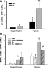
Hx/EL increases mRNA expression of HIF-1α-regulated genes, Nix and Bcl-2/adenovirus E1B-interacting protein 3 (BNIP3). Cells were treated with VEH or 7.5 U/ml EL in OxR or Hx for 8 h, and RNA was isolated. A: detection of Nix. B: detection of BNIP3. In some experiments, cells were pretreated with 10 μM SB-203580 to inhibit p38 activity. HPRT, hypoxanthine guanine phosphoribosyl transferase. aP < 0.05 vs. VEH in OxR. bP < 0.05 vs. VEH in Hx. cP < 0.05 vs. EL in OxR. %P < 0.05 vs. the same treatment without inhibitor. #Significant interaction between Hx and EL.
Fig. 7.
Fig. 7.
Hx/EL increases expression of BNIP3 protein in a p38-dependent manner. A: Hepa1c1c7 cells were treated with VEH or 7.5 U/ml EL in OxR or Hx for 12 h, and BNIP3 (25 kDa) and lamin (70 kDa) protein were detected via Western blot. B: BNIP3 and lamin intensity were measured with densitometry using ImageJ software. Values are means ± SE of 8 separate experiments. C: cells were pretreated for 2 h with DMSO (0.01% final concentration) or 10 μM SB-203580 to inhibit p38 and then treated with VEH or 7.5 U/ml EL in OxR or Hx for 12 h. BNIP3 protein was detected via Western blot. aP < 0.05 vs. VEH in OxR. bP < 0.05 vs. VEH in Hx. cP < 0.05 vs. EL in OxR.
Fig. 8.
Fig. 8.
HIF-1β-deficient HepaC4 cells are protected from the cytotoxicity of Hx/EL. Hepa1c1c7 and HepaC4 cells were treated with VEH or 7.5 U/ml EL in OxR or Hx for 24 h, and cytotoxicity was assessed. Values are means ± SE of 4 experiments in triplicate. aP < 0.05 vs. VEH in OxR. bP < 0.05 vs. EL in OxR. cP < 0.05 vs. VEH in Hx. %P < 0.05 vs. the same treatment in Hepa1c1c7 cells. #Significant interaction between treatments.
Fig. 9.
Fig. 9.
HepaC4 cells do not express BNIP3 mRNA or protein after Hx-EL cotreatment. A: HepaC4 cells were pretreated with VEH (0.01% DMSO) or 10 μM SB-203580 and then treated with VEH (PBS) or 7.5 U/ml EL in OxR or Hx for 8 h. Expression of BNIP3 and HPRT mRNA was assessed by quantitative RT-PCR. Values are means ± SE of 4 experiments in duplicate. B: Hepa1c1c7 and HepaC4 cells were treated with VEH or EL in OxR or Hx for 12 h, and whole cell lysates were probed for BNIP3 protein and lamin.
Fig. 10.
Fig. 10.
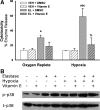
Lipid peroxidation contributes to the mechanism of Hx/EL-induced cell death in Hepa1c1c7 cells. A: Hepa1c1c7 cells were pretreated with 0.05% DMSO or 50 μM vitamin E for 30 min and then treated with VEH (PBS) or 7.5 U/ml EL in OxR or Hx for 24 h, and cytotoxicity was assessed. Values are means ± SE of 3 experiments in triplicate. B: Hepa1c1c7 cells were treated with VEH or 7.5 U/ml EL in OxR or Hx for 30 min, and whole cell extracts were probed for phosphorylated and total p38. aP < 0.05 vs. VEH in OxR. bP < 0.05 vs. VEH in Hx. cP < 0.05 vs. EL in OxR. %P < 0.05 vs. the same treatment without inhibitor.
Fig. 11.
Fig. 11.
Proposed pathway of Hx (↓Po2)/EL-induced cell death. Hx/EL increased HIF-1α accumulation and activation, leading to increased transcription of HIF-1α-directed genes BNIP3 and Nix (Figs. 5–7). Hx/EL also enhanced activation of p38 (Fig. 3). Inhibition of p38 reduced BNIP3 expression (Fig. 7) and protected cells from Hx/EL-induced cell death (Fig. 4). Deficiency in HIF-1α signaling attenuated BNIP3 expression (Fig. 9) and protected cells from Hx/EL cytotoxicity (Fig. 8). Hx/EL resulted in enhanced lipid peroxidation, which is involved in cell death (Fig. 10).

References

    1. Arteel GE, Iimuro Y, Yin M, Raleigh JA, Thurman RG. Chronic enteral ethanol treatment causes hypoxia in rat liver tissue in vivo. Hepatology 25: 920–926, 1997 - PubMed
    1. Chandel N, Maltepe E, Goldwasser E, Mathieu C, Simon M, Schumacker P. Mitochondrial reactive oxygen species trigger hypoxia-induced transcription. Proc Natl Acad Sci USA 95: 11715–11720, 1998 - PMC - PubMed
    1. Choi SM, Oh H, Park H. Microarray analyses of hypoxia-regulated genes in an aryl hydrocarbon receptor nuclear translocator (Arnt)-dependent manner. FEBS J 275: 5618–5634, 2008 - PubMed
    1. Chu W, Li X, Li C, Wan L, Shi H, Song X, Liu X, Chen X, Zhang C, Shan H, Lu Y, Yang B. TGFBR3, a potential negative regulator of TGF-β signaling, protects cardiac fibroblasts from hypoxia-induced apoptosis. J Cell Physiol 226: 2586–2594, 2011 - PubMed
    1. Conrad PW, Rust RT, Han J, Millhorn DE, Beitner-Johnson D. Selective activation of p38α and p38γ by hypoxia. Role in regulation of cyclin D1 by hypoxia in PC12 cells. J Biol Chem 274: 23570–23576, 1999 - PubMed

Publication types

MeSH terms

Substances

LinkOut - more resources