. 2012 Feb 17;110(4):569-77.
doi: 10.1161/CIRCRESAHA.111.260455. Epub 2012 Jan 5.

Shortened Ca2+ signaling refractoriness underlies cellular arrhythmogenesis in a postinfarction model of sudden cardiac death

Affiliations

Shortened Ca2+ signaling refractoriness underlies cellular arrhythmogenesis in a postinfarction model of sudden cardiac death

Andriy E Belevych et al. Circ Res. .

Abstract

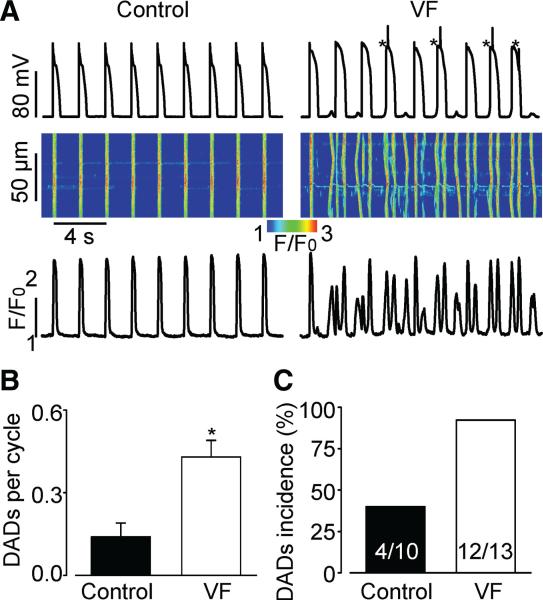
Rationale: Diastolic spontaneous Ca(2+) waves (DCWs) are recognized as important contributors to triggered arrhythmias. DCWs are thought to arise when [Ca(2+)] in sarcoplasmic reticulum ([Ca(2+)](SR)) reaches a certain threshold level, which might be reduced in cardiac disease as a consequence of sensitization of ryanodine receptors (RyR2s) to luminal Ca(2+).

Objective: We investigated the mechanisms of DCW generation in myocytes from normal and diseased hearts, using a canine model of post-myocardial infarction ventricular fibrillation (VF).

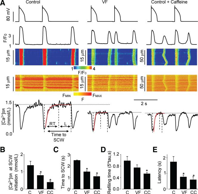
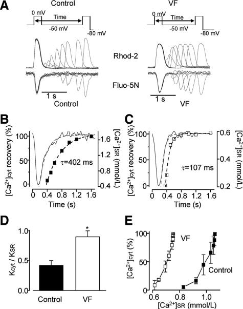
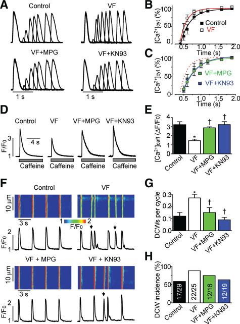
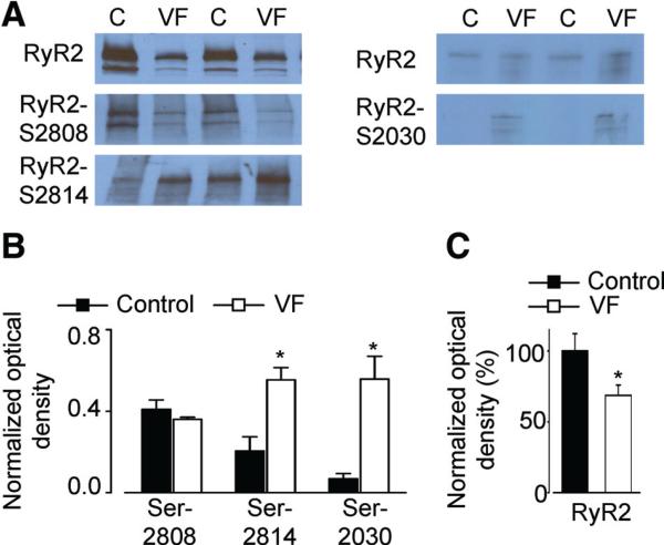
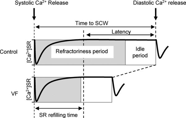
Methods and results: The frequency of DCWs, recorded during periodic pacing in the presence of a β-adrenergic receptor agonist isoproterenol, was significantly higher in VF myocytes than in normal controls. Rather than occurring immediately on reaching a final [Ca(2+)](SR), DCWs arose with a distinct time delay after attaining steady [Ca(2+)](SR) in both experimental groups. Although the rate of [Ca(2+)](SR) recovery after the SR Ca(2+) release was similar between the groups, in VF myocytes the latency to DCWs was shorter, and the [Ca(2+)](SR) at DCW initiation was lower. The restitution of depolarization-induced Ca(2+) transients, assessed by a 2-pulse protocol, was significantly faster in VF myocytes than in controls. The VF-related alterations in myocyte Ca(2+) cycling were mimicked by the RyR2 agonist, caffeine. The reducing agent, mercaptopropionylglycine, or the CaMKII inhibitor, KN93, decreased DCW frequency and normalized restitution of Ca(2+) release in VF myocytes.

Conclusions: The attainment of a certain threshold [Ca(2+)](SR) is not sufficient for the generation of DCWs. Postrelease Ca(2+) signaling refractoriness critically influences the occurrence of DCWs. Shortened Ca(2+) signaling refractoriness due to RyR2 phosphorylation and oxidation is responsible for the increased rate of DCWs observed in VF myocytes and could provide a substrate for synchronization of arrhythmogenic events at the tissue level in hearts prone to VF.


Figures

Figure 1
Figure 1. Frequency of spontaneous diastolic Ca2+ waves (DCWs) and delayed afterdepolarizations (DADs) are increased in VF myocytes
A, Representative recordings of membrane potential with corresponding line-scan images and temporal profiles of Rhod-2 fluorescence recorded in control and VF myocytes stimulated at 0.5 Hz in the presence of 100 nmol/L isoproterenol, a β-adrenergic receptor agonist. Asterisks mark action potentials triggered by DADs. B, Average frequency of DADs recorded in VF myocytes (0.43±0.06 per cycle, n=13) was significantly higher when compared with controls (0.14±0.05 per cycle, n=10, P<0.05). C, Number of cells displaying DCWs and DADs is higher in VF than in control.
Figure 2
Figure 2. Susceptibility of VF myocytes to DCWs is associated with decreased [Ca2+]SR and shortened time interval between SR Ca2+ depletion and DCW initiation
A, Action potential clamp with corresponding line-scan images and temporal profiles of Rhod-2 and Fluo-5N fluorescence recorded in myocytes from control, VF, and control treated with caffeine (0.4 mmol/L) groups. Cells were stimulated at 0.5 Hz, using a control AP waveform (upper panels). Time interval between SR Ca2+ depletion and DCW initiation can be divided into 2 periods marked by dashed vertical lines: refilling time (RT) and latency period (L, [Ca2+]SR remains constant). Post-depletion SR Ca2+ recovery was fit to exponential functions (red traces) with time constants of 256, 84, and 85 ms for control, VF, and control plus caffeine myocytes, respectively. RT was calculated as five times the fitted exponential time constant to correspond to >99% recovery. Dotted horizontal lines indicate the steady-state SR Ca2+ level at which DCWs occur in each experimental group. Bar graphs demonstrate average data for SR Ca2+ levels at initiation of DCWs (B), time after systolic SR Ca2+ depletion and DCW initiation (C), SR refilling time (D), and latency period (E) that were recorded in control (C, n=6–7), VF (n=10–11), and control plus caffeine (CC, n=7–8) groups. *P<0.05 versus control.
Figure 3
Figure 3. Ca2+ signaling refractoriness is shortened in VF myocytes
A, Upper panels show the voltage protocol used to measure recovery of the amplitude of cytosolic Ca2+ transient ([Ca2+]CYT) from SR Ca2+-dependent deactivation. Lower panels show representative recordings of Rhod-2 and Fluo-5N fluorescence during 2-pulse experiments. Time-course of [Ca2+]CYT (squares) and [Ca2+]SR (black line) recovery is shown for control (B) and VF (C) myocytes. Dashed lines represent exponential fits to the data. D, Rate of recovery of the amplitude of [Ca2+]CYT (KCYT) was normalized to the rate of recovery of [Ca2+]SR (KSR) for control (n=3) and VF (n=5) myocytes. E, [Ca2+]CYT was plotted as a function of [Ca2+]SR for control (n=3) and VF (n=5) myocytes during 2-pulse experiments described in A through C. *P<0.05 versus control.
Figure 4
Figure 4. Treatment with the reducing agent MPG or with the CaMKII inhibitor KN93 normalized recovery of cytosolic Ca2+ transient, restored the SR Ca2+ content toward control values, and significantly reduced the frequency of DCWs recorded in VF myocytes
A, Representative recordings of cytosolic Ca2+ transients imaged using Fluo-4FF during 2-pulse experiments (shown in Figure 3) in voltage-clamped control and VF myocytes and VF myocytes treated with either 0.75 mmol/L MPG or 1 μmol/L KN93. B and C, Average amplitude of the cytosolic Ca2+ transients presented as a function of interpulse time. The recovery of the amplitude was fitted to a mono exponential function with time constants of 287±16 ms in control (n=7), 197±28 ms in VF untreated (n=3–4), 342±33 ms in VF myocytes treated with MPG (n=4–5), and 380±40 ms in VF treated with KN93 (n=4–5). D, Representative recordings of cytosolic Ca2+ transients induced by 10 mmol/L caffeine imaged with Fluo-4FF in voltage-clamped control and VF myocytes. E, average amplitude of caffeine-induced Ca2+ transients in control (n=8) and VF (n=8) myocytes, and VF myocytes treated with either 0.75 mmol/L MPG (n=6) or 1 μmol/L KN93 (n=4). F, Representative line-scan images and temporal profiles of Rhod-2 fluorescence recorded in control and VF myocytes, and in VF myocytes treated with either 0.75 mmol/L MPG, a reducing agent, or 1 μmol/L KN-93, a CaMKII inhibitor. Cells were field-stimulated at 0.3 Hz. G, Frequency of DCWs (marked with arrows in F) was calculated for control (n=29) and VF myocytes (n=25) and VF myocytes treated with either MPG (n=19) or KN-93 (n=16). H, Proportion of cells displaying DCWs in each experimental group. All data presented in this figure were obtained in the presence of 100 nmol/L isoproterenol, a β-adrenergic receptor agonist. *P<0.05 versus control; †P<0.05 versus VF untreated.
Figure 5
Figure 5. Phosphorylation levels of RyR2 are altered in VF
A, Representative Western blots showing phosphorylation of RyR2s at Ser-2030 and Ser-2808 (PKA-dependent) and Ser-2814 (CaMKII-dependent) phosphorylation sites in control and in VF myocytes measured with phospho-specific antibodies. B, Data pooled for Ser 2030 (n=5), Ser-2808 (n=7), and Ser-2814 (n=4) experiments, respectively. C, Average data show signifi-cant decrease in the amount of RyR2 in VF (n=8) compared with control (n=8). *P<0.05 versus control.
Figure 6
Figure 6. Schematic representation of the time delay between systolic SR Ca2+ depletion and DCW in control and VF myocytes
The time delay between systolic SR Ca2+ depletion and DCW is comprised of a period of refilling of the SR Ca2+ store and a latency period during which [Ca2+]SR remains constant. The SR Ca2+ refilling time was not significantly different between control and VF myocytes, whereas latency was significantly shorter in VF myocytes (Figure 2). The refractory period reflects the time required for recovery of RyR2s from the SR Ca2+-dependent deactivation. In control myocytes, the refractory period determined in 2-pulse experiments was 2 times longer than the SR Ca2+ refilling time (Figure 3). In VF myocytes the refractory period was approximately equal to the SR Ca2+ refilling time (Figure 3). In both control and VF groups, the refractory period is followed by an idle period, during which stochastic activation of the recovered SR Ca2+ release sites triggers a DCW.

References

    1. Lloyd-Jones D, Adams RJ, Brown TM, Carnethon M, Dai S, De Simone G, Ferguson TB, Ford E, Furie K, Gillespie C, Go A, Greenlund K, Haase N, Hailpern S, Ho PM, Howard V, Kissela B, Kittner S, Lackland D, Lisabeth L, Marelli A, McDermott MM, Meigs J, Mozaffarian D, Mussolino M, Nichol G, Roger VL, Rosamond W, Sacco R, Sorlie P, Roger VL, Thom T, Wasserthiel-Smoller S, Wong ND, Wylie-Rosett J. Heart disease and stroke statistics: 2010 update: a report from the American Heart Association. Circulation. 2010;121:e46–e215. - PubMed
    1. Bunch TJ, Hohnloser SH, Gersh BJ. Mechanisms of sudden cardiac death in myocardial infarction survivors: insights from the randomized trials of implantable cardioverter-defibrillators. Circulation. 2007;115:2451–2457. - PubMed
    1. Ter Keurs HE, Boyden PA. Calcium and arrhythmogenesis. Physiol Rev. 2007;87:457–506. - PMC - PubMed
    1. Laurita KR, Rosenbaum DS. Mechanisms and potential therapeutic targets for ventricular arrhythmias associated with impaired cardiac calcium cycling. J Mol Cell Cardiol. 2008;44:31–43. - PMC - PubMed
    1. Weiss JN, Nivala M, Garfinkel A, Qu Z. Alternans and arrhythmias: from cell to heart. Circ Res. 2011;108:98–112. - PMC - PubMed

Publication types

MeSH terms