. 2012 Feb 17;110(4):551-9.
doi: 10.1161/CIRCRESAHA.111.255927. Epub 2012 Jan 5.

Left-right symmetry breaking in tissue morphogenesis via cytoskeletal mechanics

Affiliations

Left-right symmetry breaking in tissue morphogenesis via cytoskeletal mechanics

Ting-Hsuan Chen et al. Circ Res. .

Abstract

Rationale: Left-right (LR) asymmetry is ubiquitous in animal development. Cytoskeletal chirality was recently reported to specify LR asymmetry in embryogenesis, suggesting that LR asymmetry in tissue morphogenesis is coordinated by single- or multi-cell organizers. Thus, to organize LR asymmetry at multiscale levels of morphogenesis, cells with chirality must also be present in adequate numbers. However, observation of LR asymmetry is rarely reported in cultured cells.

Objectives: Using cultured vascular mesenchymal cells, we tested whether LR asymmetry occurs at the single cell level and in self-organized multicellular structures.

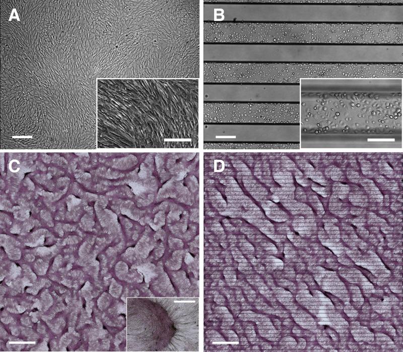
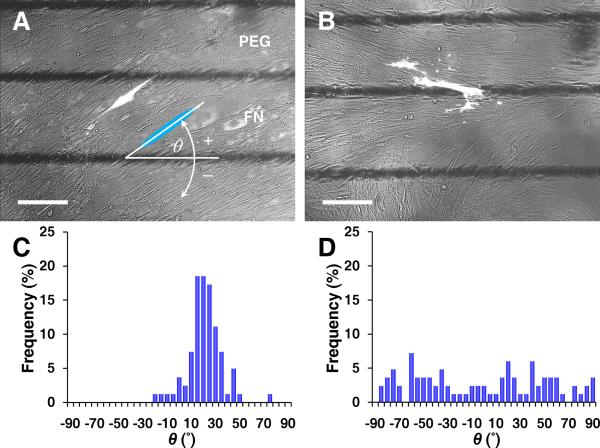
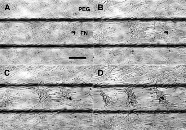
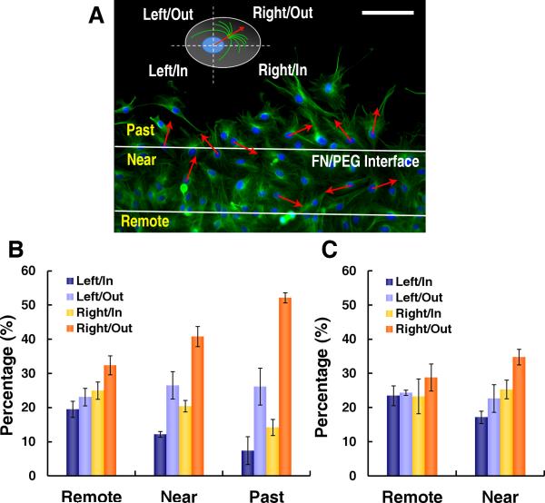
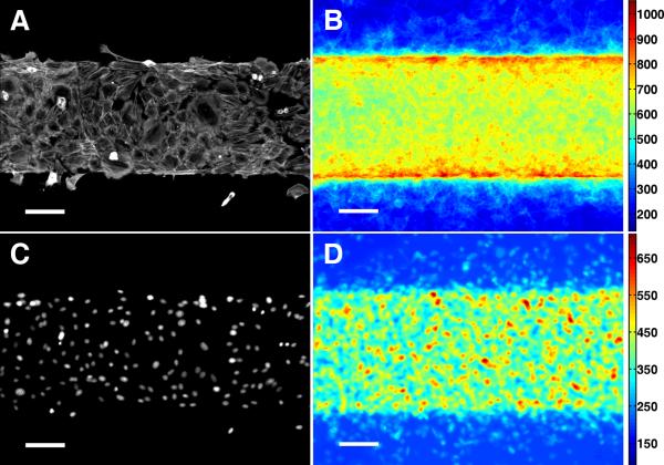
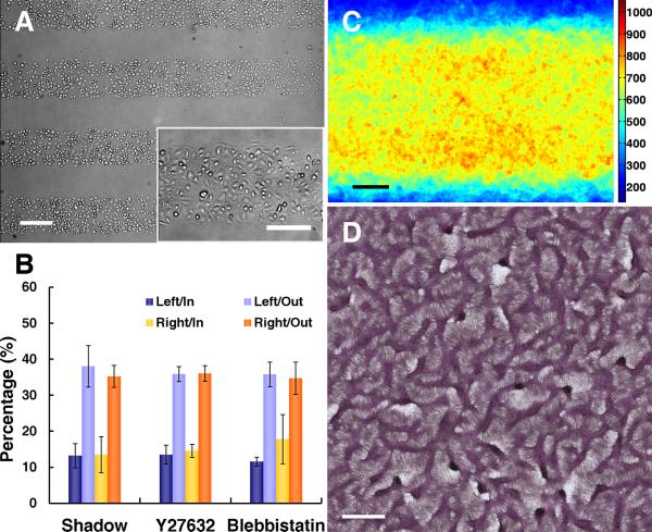
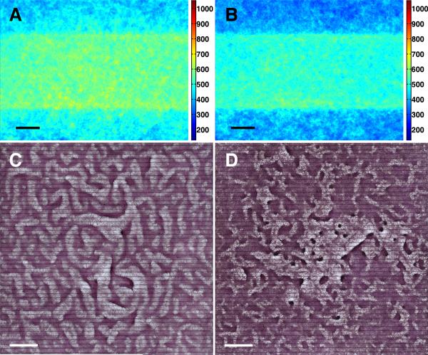
Methods and results: Using micropatterning, immunofluorescence revealed that adult vascular cells polarized rightward and accumulated stress fibers at an unbiased mechanical interface between adhesive and nonadhesive substrates. Green fluorescent protein transfection revealed that the cells each turned rightward at the interface, aligning into a coherent orientation at 20° relative to the interface axis at confluence. During the subsequent aggregation stage, time-lapse videomicroscopy showed that cells migrated along the same 20° angle into neighboring aggregates, resulting in a macroscale structure with LR asymmetry as parallel, diagonal stripes evenly spaced throughout the culture. Removal of substrate interface by shadow mask-plating, or inhibition of Rho kinase or nonmuscle myosin attenuated stress fiber accumulation and abrogated LR asymmetry of both single-cell polarity and multicellular coherence, suggesting that the interface triggers asymmetry via cytoskeletal mechanics. Examination of other cell types suggests that LR asymmetry is cell-type specific.

Conclusions: Our results show that adult stem cells retain inherent LR asymmetry that elicits de novo macroscale tissue morphogenesis, indicating that mechanical induction is required for cellular LR specification.


Figures

Figure 1
Figure 1. LR asymmetry in pattern formation by vascular mesenchymal cells
Phase contrast microscopy of VMCs on (A) conventional plastic substrate at confluence and (B) FN/PEG substrates (interfaces identified by black titanium lines) showing preferential attachment to FN domain immediately after plating. Scale bar, 300 μm and 200 μm (inset). After 10-14 days, development of regularly spaced aggregates (C) in a labyrinthine configuration and (D) in a stripe pattern along principal diagonal axis in bright field (multicellular ridges stained with hematoxylin). Insets: higher magnification images of multicellular aggregates. Scale bar, 2 mm and 300 μm (inset).
Figure 2
Figure 2. Early stage of coherent single-cell orientation perpendicular to the axis of diagonal ridges
Fluorescence microscopy of GFP-transfected VMCs on (A) FN/PEG substrates showing coherent individual cell orientation, relative to the interface axis (black lines) and (B) control substrate as homogeneous FN. Scale bar, 200 μm. Histogram of θ for (C) FN/PEG, showing convergence to 19 ± 14° (n = 81 cells; day 5; mean ± s.d.) and (D) control substrate, showing non-convergence (n = 83 cells; day 5).
Figure 3
Figure 3. Cell migration toward aggregates directed by the coherent orientation
Time-lapse videomicroscopic images at (A) t = 0, (B) t = 150, (C) t = 300, and (D) t = 625 min. Scale bar, 200 μm.
Figure 4
Figure 4. Numerical simulations showing that the anisotropic migration leads to parallel ridges with asymmetric alignment
A, Schematic of coefficients, b1 and b2, for principal directions of anisotropic migration. Simulation results for n(x, y) with darker areas representing higher density yielded (B) a labyrinthine pattern at steady state for isotropic migration (b1 = b2 = 1) and (C) an asymmetric pattern for anisotropic migration (b1 = 1, b2 = 10-6). Model parameters: D = 0.005, γ = 20000, k = 0.28, c = 0.01, e = 0.02, Dn = 0.06, χ0 = 0.06, kn = 1, θ = 20°, r = 322, t* = 1 (total time).
Figure 5
Figure 5. LR polarity at FN/PEG interface
A, Immunofluorescence microscopy of α-tubulin in VMCs on FN/PEG with polarity defined based on MTOC orientation relative to nuclear centroid and interface for cells past, near, and 120 μm remote from interface. Scale bar, 100 μm. VMC polarity (B) 6 hours after plating (n = 3 experiments; > 370 cells each; mean ± s.d.) and (C) 2 hours after plating (n = 3 experiments; > 260 cells each; mean ± s.d.).
Figure 6
Figure 6. Stress fiber accumulation at FN/PEG interface
Immunofluorescence microscopy of NMM-IIa in VMCs on FN/PEG substrate shown as (A) a single image and (B) stacked images (n = 45). Immunofluorescence microscopy of nuclei of VMCs on FN/PEG substrate shown as (C) a single image and (D) stacked images (n = 45). Scale bar, 100 μm.
Figure 7
Figure 7. Abrogation of LR polarity and LR alignment of multicellular ridges with removal of substrate interfaces
A, Microscopic images after shadow-mask plating show plating limited to rectangular domains. Scale bar, 300 μm and 200 μm (inset). B, Polarity of VMCs at the edges of cell sheets shadow-plated onto uniform FN (n = 3 experiments; > 175 cells each; mean ± s.d.) and VMCs near the FN/PEG interface with Y27632 (n = 3; > 135 cells each) or blebbistatin (n = 3; > 150 cells each) inhibition. C, Stacked images of NMM-IIa immunofluorescence after shadow-plating on FN (n = 40). Scale bar, 100 μm. D, Multicellular patterns by hematoxylin staining after shadow-plating on FN substrate. Scale bar, 2 mm.
Figure 8
Figure 8. Abrogation of LR alignment of multicellular ridges with stress fiber inhibition
Stacked images of NMM-IIa immunofluorescence (A) with Y27632 on FN/PEG substrate (n = 60), and (B) with blebbistatin on FN/PEG substrate (n= 45). Scale bar, 100 μm. Multicellular patterns by hematoxylin staining (C) with Y27632 on FN/PEG substrate, and (D) with blebbistatin on FN/PEG substrate. Scale bar, 2 mm.

References

    1. Okumura T, Utsuno H, Kuroda J, Gittenberger E, Asami T, Matsuno K. The Development and Evolution of Left-Right Asymmetry in Invertebrates: Lessons From Drosophila and Snails. Dev Dyn. 2008;237:3497–3515. - PubMed
    1. Aw S, Levin M. Is left-right asymmetry a form of planar cell polarity? Development. 2009;136:355–366. - PMC - PubMed
    1. Hashimoto M, Shinohara K, Wang J, Ikeuchi S, Yoshiba S, Meno C, Nonaka S, Takada S, Hatta K, Wynshaw-Boris A, Hamada H. Planar polarization of node cells determines the rotational axis of node cilia. Nat Cell Biol. 2010;12:170–176. - PubMed
    1. Song H, Hu J, Chen W, Elliott G, Andre P, Gao B, Yang Y. Planar cell polarity breaks bilateral symmetry by controlling ciliary positioning. Nature. 2010;466:378–382. - PMC - PubMed
    1. Danilchik MV, Brown EE, Riegert K. Intrinsic chiral properties of the Xenopus egg cortex: an early indicator of left-right asymmetry? Development. 2006;133:4517–4526. - PubMed

Publication types

MeSH terms

Substances

LinkOut - more resources