Review
. 2012 Jul;18(7):1378-88.
doi: 10.1002/ibd.22859. Epub 2012 Jan 4.

Implications of protein post-translational modifications in IBD

Affiliations
Review

Implications of protein post-translational modifications in IBD

Stefan F Ehrentraut et al. Inflamm Bowel Dis. 2012 Jul.

Abstract

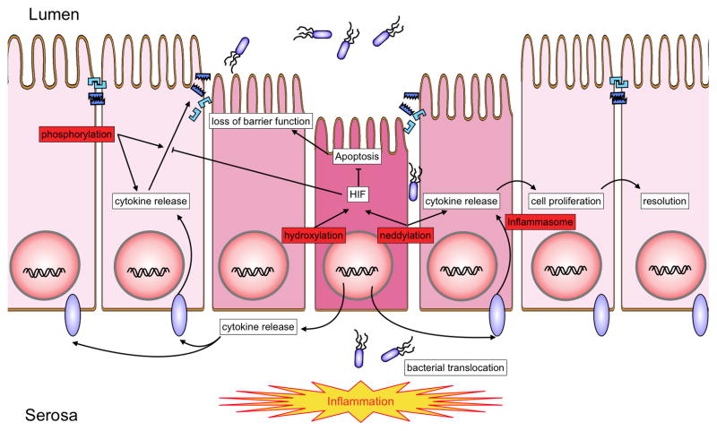
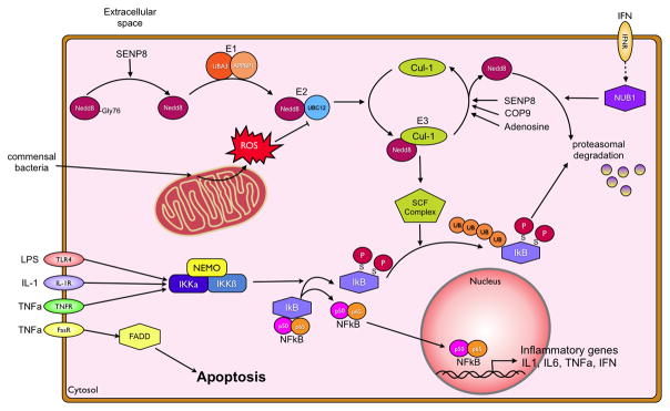
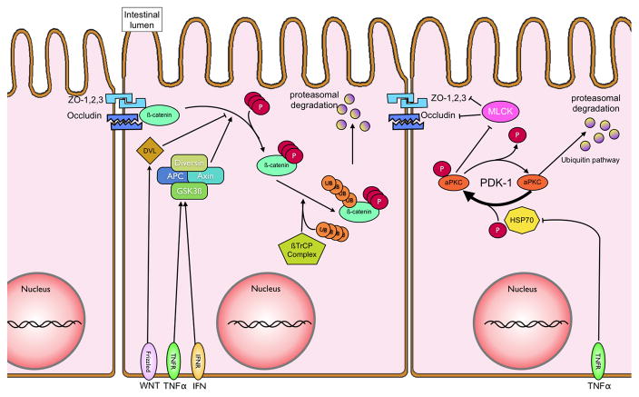
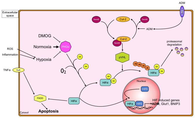
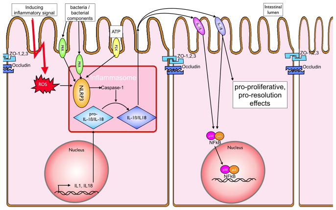
In recent years our understanding of the pathogenesis of inflammatory bowel disease (IBD) has greatly increased. Hallmarks of IBD include loss of intestinal barrier function, increased cytokine production, and failed resolution of tissue damage. Lasting treatments are still lacking and, therefore, a better understanding of the underlying molecular mechanisms is necessary to design novel therapeutic approaches. Apart from transcriptional and posttranscriptional regulation of relevant genes, mammals have evolved a complex and efficient series of mechanisms to rapidly modify newly made proteins for the purposes of signaling and adaptation. These posttranslational protein modifications include, among others, phosphorylation, hydroxylation, neddylation, and cytokine cleavage by the inflammasome. This review focuses on our current understanding of posttranslational protein modifications with a particular focus on their relevance to IBD pathogenesis.


Figures

Figure 1
Figure 1. Overview of posttranslational protein modifications relevant to IBD
A number of posttranslational protein modifications relevant to IBD (highlighted in red). These pathways include phosphorylation, neddylation hydroxylation and cleavage of cytokine precursor forms by the inflammasome. The interplay of these rapid response mechanisms enables rapid adaptation to incoming inflammatory signals. Cytokine induced barrier breakdown allows for bacterial translocation to the basal aspect of intestinal epithelial cells. Bacterial antigens and endogenous danger signals are recognized by the adaptive and innate immune system, triggering a variety of reactions including apoptosis, increased cytokine release, loss of tight junctional proteins and barrier breakdown.
Figure 2
Figure 2. Phosphorylation and neddylation pathways
Recognition of pro-inflammatory stimuli, presence of microbial cell wall components, secreted TNFα or various other cytokines, through their respective receptors triggers a pro-inflammatory first response of the IEC. The NFκB pathway is activated through the phosphorylation of IκB by the Iκ-kinases α and β. This phosphorylation allows for its recognition by the neddylated Skp-Cullin-F-Box (SCF) complex, polyubiquitination and subsequent proteasomal degradation. Neddylation of Cullins is achieved through a multienzyme process, conjugating a NEDD8 moiety to the target protein. NEDD8, in order to be conjugated has to be processed from its pro-form to the mature form by the isopeptidase SENP8. The same protein, in addition to the COP9 signalosome removes NEDD8 from Cullins. Neddylated Cullins are integrated into SCF complex and activate it to enable SCF complex mediated ubiquitination. The NFκB heterodimer can then translocate to the nucleus and bind to the promoter regions of various pro- and anti-inflammatory cytokines, including TNFα, IL-1β and interferon-γ. This can further promote inflammation, e.g. by TNFα induced apoptosis through the FADD pathway, and barrier breakdown, but also abrogate the incoming stimulus by induction of NUB1 through the interferon receptor. Anti-inflammatory mechanisms include the induction of deneddylation of Cullin-proteins through adenosine and ROS mediated inhibition of the E2 ligase NEDD8/UBC12.
Figure 3
Figure 3. Intestinal barrier regulation in iflammation
Following NFκB activation, various cytokines, including TNFα, are secreted in a paracrine fashion. Recognition of TNFα by its surface receptor TNFR1, inhibits heat shock protein 70 (HSP70). HSP70 mediates the re/-phosphorylation of atypical Protein Kinase C (aPKC) by serving as a chaperoning agent for the unphosphorylated aPKC. Phosphorylated aPKC inhibits Myosin light chain kinases, which prevent full maturation of tight junctional proteins (zonula occludens 1,2,3 and occludins) leading to decreased transepithelial resistance and loss of barrier function. Tight junctional integrity also relies on the interaction between β-catenin and the cytoskeleton. TNF-α and interferon-gamma, secreted during inflammation, can activate a multienzyme complex comprised of diversin, adenomatous polyposis coli (APC), Axin and glycogen synthase 3b (GSK3β). The GSK3β subunit phosphorylates β-catenin, allowing for its recognition and ubiquitination by the β-TrCP/E3 ligase. Ubiquinated β-catenin is subsequently proteasomally degraded, and thus unavailable for enhancing barrier function.
Figure 4
Figure 4. Regulation of hypoxia-inducible factor by hydroxylation
Under normoxic conditions, the HIFα subunit is hydroxylated through prolylhydroxylases 1–3 (PHDs) or the Factor-inhibiting-HIF (FIH). Hydroxylated HIF is recognized by the von Hippel-Lindau (pVHL) protein, which in its activated state contains a neddylated Culllin-2 subunit. This leads to ubiquitination of the HIF1α subunit and its degradation by the proteasome. During inflammation, local oxygen is limited due to increased metabolism and also due to neutrophil derived reactive oxygen species. This hypoxic environment renders PHDs impotent, as does pharmacological inhibition with DMOG, preventing ubiquitination of HIFα by pVHL. Non-ubiquitinated HIFα translocates to the nucleus, binds to the beta-subunit and the heterodimer functions as a transcription factor of a variety of genes, including adrenomedullin (ADM). Furthermore, HIFα inhibits Fas-associated-death-domain (FADD) induced apoptosis, thereby increasing intestinal barrier function. The autocrine release of ADM can, through the ADM receptor-mediated deneddylation of Cul2, serve as a negative feedback mechanism.
Figure 5
Figure 5. The inflammasome and IBD
Activation of the NLRP3 inflammasome by either cell surface (e.g. TLR4 via the TRIF pathway) or intracellular (e.g. TLR9 or NOD-receptors) pattern recognition receptors leads to the cleavage of the IL-1β and IL-18 precursors to their mature form. This activation is achieved through pathogen associated molecular patterns (PAMPs) and also through endogenous danger associated molecular patterns (DAMPs) such as extracellular ATP (via the P2X7 receptor) or intracellular reactive oxygen species (ROS). IL-1β and IL-18, when secreted, bind to their respective receptors and activate the NFκB pathway. In addition, there are possible anti-inflammatory influences attributed to IL-18 and enhanced cell proliferation, thereby leading to more rapid resolution of intestinal barrier breakdown.

References

    1. Xavier RJ, Podolsky DK. Unravelling the pathogenesis of inflammatory bowel disease. Nature. 2007;448:427–434. - PubMed
    1. Marks DJ, Rahman FZ, Sewell GW, et al. Crohn’s disease: an immune deficiency state. Clin Rev Allergy Immunol. 2010;38:20–31. - PMC - PubMed
    1. Barnes PJ, Karin M. Nuclear factor-κB - A pivitol transcription factor in chronic inflammatory disease. N Engl J Med. 1997;336:1066–1071. - PubMed
    1. Wojcik C, Di Napoli M. Ubiquitin-proteasome system and proteasome inhibition: new strategies in stroke therapy. Stroke. 2004;35:1506–1518. Epub 2004 Apr 1529. - PubMed
    1. Chen Z, Hagler J, Palombella VJ, et al. Signal-induced site-specific phosphorylation targets I kappa B alpha to the ubiquitin-proteasome pathway. Genes Dev. 1995;9:1586–1597. - PubMed

Publication types

MeSH terms