. 2012 Mar 2;287(10):7110-20.
doi: 10.1074/jbc.M111.316075. Epub 2012 Jan 5.

Structural insights into apoptotic DNA degradation by CED-3 protease suppressor-6 (CPS-6) from Caenorhabditis elegans

Affiliations

Structural insights into apoptotic DNA degradation by CED-3 protease suppressor-6 (CPS-6) from Caenorhabditis elegans

Jason L J Lin et al. J Biol Chem. .

Abstract

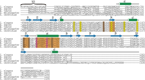
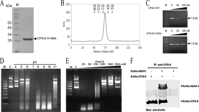
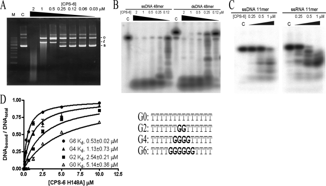
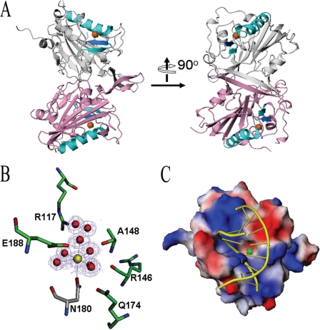
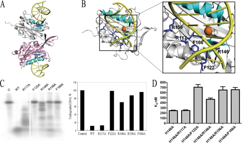
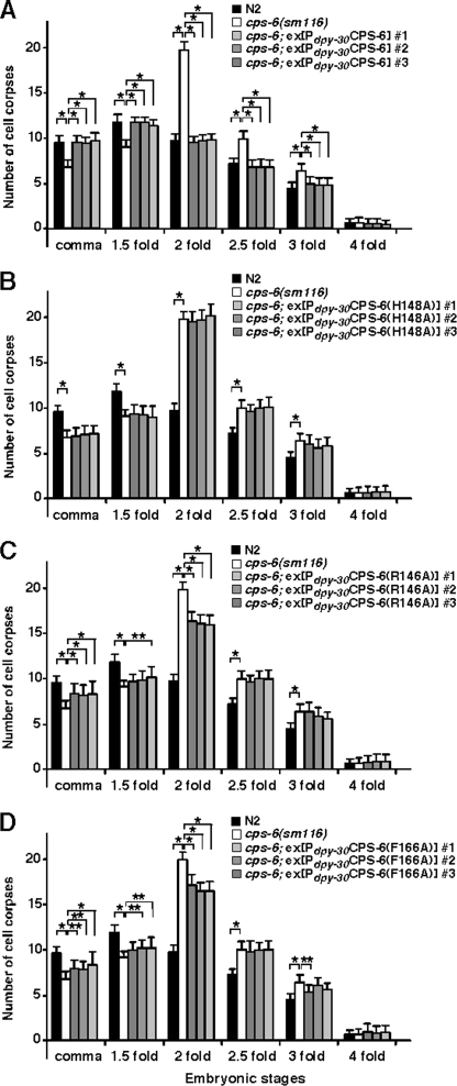
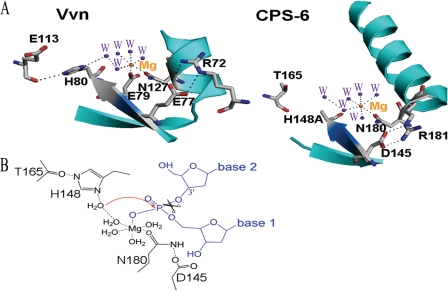
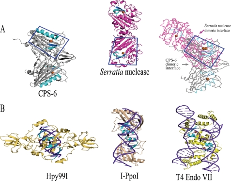
Endonuclease G (EndoG) is a mitochondrial protein that traverses to the nucleus and participates in chromosomal DNA degradation during apoptosis in yeast, worms, flies, and mammals. However, it remains unclear how EndoG binds and digests DNA. Here we show that the Caenorhabditis elegans CPS-6, a homolog of EndoG, is a homodimeric Mg(2+)-dependent nuclease, binding preferentially to G-tract DNA in the optimum low salt buffer at pH 7. The crystal structure of CPS-6 was determined at 1.8 Å resolution, revealing a mixed αβ topology with the two ββα-metal finger nuclease motifs located distantly at the two sides of the dimeric enzyme. A structural model of the CPS-6-DNA complex suggested a positively charged DNA-binding groove near the Mg(2+)-bound active site. Mutations of four aromatic and basic residues: Phe(122), Arg(146), Arg(156), and Phe(166), in the protein-DNA interface significantly reduced the DNA binding and cleavage activity of CPS-6, confirming that these residues are critical for CPS-6-DNA interactions. In vivo transformation rescue experiments further showed that the reduced DNase activity of CPS-6 mutants was positively correlated with its diminished cell killing activity in C. elegans. Taken together, these biochemical, structural, mutagenesis, and in vivo data reveal a molecular basis of how CPS-6 binds and hydrolyzes DNA to promote cell death.


Figures

FIGURE 1.
FIGURE 1.
Sequence alignment of CPS-6/EndoG. Sequences of CPS-6/EndoG from C. elegans, Homo sapiens, Bos taurus, D. melanogaster, and Saccharomyces cerevisiae are aligned and listed. CPS-6 shares high sequence identities of 50, 49, 55, and 39% with human, bovine, fruit fly, and yeast EndoG, respectively. The amino acid residues found in H. sapiens, B. taurus, and D. melanogaster important for metal ion coordination and DNA binding are colored in pink and brown, respectively (22, 23, 26, 30). The ββα-metal finger motif (β4-β5-α4) is marked in orange, and the conserved 145DRGH148 sequence is located in β4. The residues that are likely involved in DNA binding and that were subjected to site-directed mutagenesis in this study are colored in yellow. The secondary structures derived from the crystal structure of CPS-6 are depicted as green cylinders for α-helices and blue arrows for β-strands. MLS, mitochondrial localization sequence.
FIGURE 2.
FIGURE 2.
The recombinant CPS-6 is a functional homodimeric nuclease. A, the purity of the recombinant CPS-6 (63–305) H148A mutant was assayed by 12% SDS-PAGE. B, size exclusion chromatographic profile of CPS-6 H148A mutant showing that the protein was eluted at 74 ml, indicating a dimeric conformation. The protein markers used here are ferritin (440 kDa), catalase (232 kDa), bovine serum albumin (67 kDa), ovalbumin (44 kDa), and carbonic anhydrase (29 kDa). C, wild-type CPS-6 digests 1.6-kb linear DNA fragments (upper panel) in a concentration-dependent manner, whereas H148A has no nuclease activity (lower panel). Lane M is a DNA marker. D, CPS-6 digests plasmid DNA most efficiently at pH 7. pET28 plasmid DNA (25 ng) was incubated for 1 h at 37 °C with 2 μm CPS-6 in reaction buffer containing 25 mm NaCl, 2 mm MgCl2, and 2 mm DTT with pH values ranging from 4 to 11. Lane M is a DNA marker, and lane C is a control in the absence of CPS-6. E, CPS-6 digests plasmid DNA more efficiently at low salt concentrations (25–100 mm NaCl). F, the immunoprecipitation experiment shows that the His6-tagged CPS-6 (H148A mutant) interacts directly with the His-tagged WAH-1.
FIGURE 3.
FIGURE 3.
CPS-6 digests both DNA and RNA with a preference for G-track DNA. A, CPS-6 digests pET28 plasmid DNA (25 ng) in a concentration-dependent manner (0.03–2 μm). Lane M is a DNA marker. B, CPS-6 digests both 5′-end 32P-labeled single-stranded DNA (48 nucleotides) and double-stranded DNA (48 bp). C, CPS-6 digests 11-mer ssRNA (5′-end 32P-labeled 5′-AACCUUACAAC-3′) and 11-mer ssDNA (5′-end 32P-labeled 5′-AACCTTACAAC-3′) substrate. All of the reactions in A–C were carried out in the buffer of 10 mm HEPES, pH 7.0, 100 mm NaCl, 2 mm MgCl2, and 2 mm DTT for 1 h at 37 °C. Lanes C are controls in the absence of CPS-6. D, the binding constants between CPS-6 H148A mutant and the 14-nucleotide 5′-end 32P-labeled ssDNA containing different lengths of poly(dG) were measured by nitrocellulose filter binding assays. The dissociation constants between CPS-6 H148A and ssDNA containing zero (open triangle), two (circle), four (triangle), and six G (closed rectangle) are 5.14 ± 0.36, 2.54 ± 0.21, 1.13 ± 0.73, and 0.53 ± 0.02 μm, respectively. The error bars are generated from three independent experiments.
FIGURE 4.
FIGURE 4.
Crystal structure of the Mg2+-bound CPS-6 H148A mutant. A, side view of the overall crystal structure of the dimeric CPS-6 (residues 63–305, H148A mutant) that was determined at 1.8 Å resolution (Protein Data Bank code 3S5B). The two protomers are displayed in pink and gray, respectively. The ββα-metal finger motifs are displayed in cyan with a Mg2+ ion (orange sphere) bound at the active site. B, omit map illustration for the active site of CPS-6 with the Mg2+ and the coordinated water molecules omitted for the map calculation (contoured at 3.0 σ). C, top view of the molecular model of CPS-6 bound with DNA. This model was constructed by superimposition of the ββα-metal finger motif in CPS-6 (residues 144–155 and 170–182) with that of the Vvn-DNA complex (Protein Data Bank code 1OUP) to determine where the DNA (GCGATCGC) is bound on CPS-6. A positively charged surface (in blue) near the active site of CPS-6 interacts well with the DNA phosphate backbone. The Mg2+ ion in the active site is displayed as a green ball.
FIGURE 5.
FIGURE 5.
Identification of the residues involved in DNA binding in CPS-6. A, side view of a model of CPS-6 homodimer bound with two DNA molecules. The ββα-metal motif is colored in cyan, and the Mg2+ ion is in orange. The dsDNA molecules are colored in yellow. B, the top view of CPS-6-DNA complex model suggests that several basic and aromatic amino acid residues (in blue) are located closely to the DNA backbones, likely involved in protein-DNA interactions, including Arg117, Phe122, Arg146, Arg156, and Phe166. C, the DNA digestion assays show that a mutation at Phe122, Arg146, Arg156, and Phe166, but not at Arg117, greatly reduced the DNase activity of CPS-6, suggesting that these residues are involved in DNA binding and/or digestion. The integrated density values (shown as percentages, as normalized using the control DNA as 100%) for the undigested full-length 5′-end 32P-labeled ssDNA used in this study are summarized as a histogram. D, the dissociation constants between CPS-6 mutants and DNA (5′-end 32P-labeled 48-nt ssDNA) were measured by filter binding assays. The dissociation constants are shown in the histogram: 245 ± 18 nm for H148A, 249 ± 20 for H148A/R117A, 707 ± 88 nm for H148A/F122A, 468 ± 42 nm for H148A/R146A, 652 ± 111 nm for H148A/R156A, and 658 ± 74 nm for H148A/F166A.
FIGURE 6.
FIGURE 6.
The cell death assay in C. elegans. Transgenic cps-6(sm116) animals expressing wild-type CPS-6 (A), CPS-6(H148A) (B), CPS-6(R146A) (C), or CPS-6(F166A) (D) under the control of the dpy-30 promoter were generated, and the numbers of cell corpses were scored. For each construct, the data were collected from three independent transgenic lines. The stages of transgenic embryos examined were: comma and 1.5-, 2-, 2.5-, 3-, and 4-fold. The y axis represents the average number of cell corpses scored, and the error bars show the standard deviations. Fifteen embryos were counted for each developmental stage. The significance of differences were determined by two-way analysis of variance, followed by Bonferroni comparison. *, p < 0.001; **, p < 0.05. All other points had p values > 0.05.
FIGURE 7.
FIGURE 7.
The active site and proposed catalytic mechanism for CPS-6. A, the active site of CPS-6 shares a similar conformational arrangement with that of Vvn. The ββα-metal finger motif is colored in cyan, with the conserved 145DRGH148 sequence (and the corresponding 77EWEH80 sequence in Vvn) displayed in marine blue. B, schematic diagram of the proposed DNA hydrolysis mechanism by CPS-6. His148 acts as a general base to activate a water molecule, which in turn makes an in-line attack on the scissile phosphate. The magnesium ion stabilizes the phosphoanion transition state, and the Mg2+-bound water molecule functions as a general acid to provide a proton to the 3′-oxygen leaving group.
FIGURE 8.
FIGURE 8.
Structural comparison of nonspecific and site-specific dimeric ββα-metal finger nucleases. A, the overall folds of the two nonspecific nucleases, CPS-6 and Serratia nuclease, are similar. However, the dimeric interfaces are located in different regions as revealed by the superimposition of one protomer of the two proteins (boxed in the right panel). B, the crystal structures of the three site-specific endonucleases Hpy99I, I-PpoI, and T4 Endo VII in complex with their DNA substrates show that the two ββα-metal motifs are positioned and oriented next to the DNA sugar-phosphate backbones. The 2-fold symmetry (displayed as an oval) of the dimeric proteins coincides roughly with the 2-fold axis of the DNA substrates.

Similar articles

Cited by

References

    1. Ruiz-Carrillo A., Renaud J. (1987) Endonuclease G. A (dG)n × (dC)n-specific DNase from higher eukaryotes. EMBO J. 6, 401–407 - PMC - PubMed
    1. Cummings O. W., King T. C., Holden J. A., Low R. L. (1987) Purification and characterization of the potent endonuclease in extracts of bovine heart mitochondria. J. Biol. Chem. 262, 2005–2015 - PubMed
    1. Low R. L. (2003) Mitochondrial endonuclease G function in apoptosis and mtDNA metabolism. A historical perspective. Mitochondrion 2, 225–236 - PubMed
    1. Parrish J., Li L., Klotz K., Ledwich D., Wang X., Xue D. (2001) Mitochondrial endonuclease G is important for apoptosis in C. elegans. Nature 412, 90–94 - PubMed
    1. Li L. Y., Luo X., Wang X. (2001) Endonuclease G is an apoptotic DNase when released from mitochondria. Nature 412, 95–99 - PubMed

Publication types

MeSH terms

Associated data

LinkOut - more resources