. 2012 Mar 2;287(10):7399-410.
doi: 10.1074/jbc.M111.296608. Epub 2012 Jan 5.

Induction of DNA damage signaling upon Rift Valley fever virus infection results in cell cycle arrest and increased viral replication

Affiliations

Induction of DNA damage signaling upon Rift Valley fever virus infection results in cell cycle arrest and increased viral replication

Alan Baer et al. J Biol Chem. .

Abstract

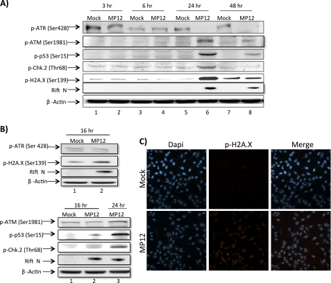
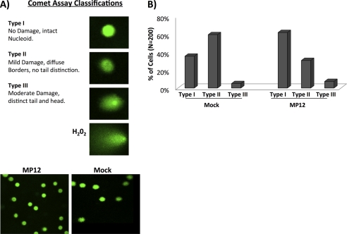
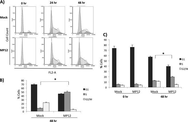
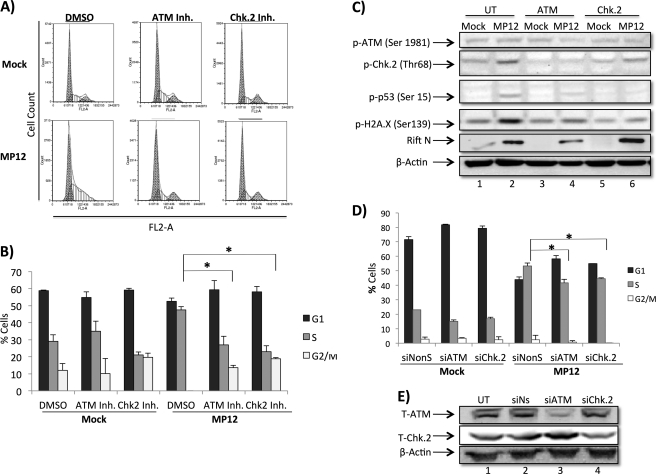
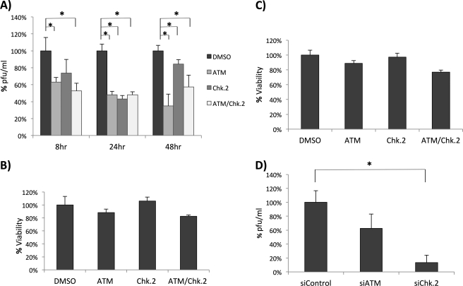
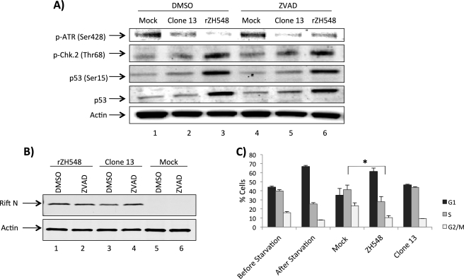
Rift Valley fever virus (RVFV) is a highly pathogenic arthropod-borne virus infecting a wide range of vertebrate hosts. Of particular interest is the nonstructural NSs protein, which forms large filamentous fibril bundles in the nucleus. Past studies have shown NSs to be a multifaceted protein important for virulence through modulation of the interferon response as well acting as a general inhibitor of transcription. Here we investigated the regulation of the DNA damage signaling cascades by RVFV infection and found virally inducted phosphorylation of the classical DNA damage signaling proteins, ataxia-telangiectasia mutated (ATM) (Ser-1981), Chk.2 (Thr-68), H2A.X (Ser-139), and p53 (Ser-15). In contrast, ataxia-telangiectasia mutated and Rad3-related kinase (ATR) (Ser-428) phosphorylation was decreased following RVFV infection. Importantly, both the attenuated vaccine strain MP12 and the fully virulent strain ZH548 showed strong parallels in their up-regulation of the ATM arm of the DNA damage response and in the down-regulation of the ATR pathway. The increase in DNA damage signaling proteins did not result from gross DNA damage as no increase in DNA damage was observed following infection. Rather the DNA damage signaling was found to be dependent on the viral protein NSs, as an NSs mutant virus was not found to induce the equivalent signaling pathways. RVFV MP12-infected cells also displayed an S phase arrest that was found to be dependent on NSs expression. Use of ATM and Chk.2 inhibitors resulted in a marked decrease in S phase arrest as well as viral production. These results indicate that RVFV NSs induces DNA damage signaling pathways that are beneficial for viral replication.


Figures

FIGURE 1.
FIGURE 1.
DNA damage signaling is induced following RVFV MP12 infection. A, HSAECs were mock-infected or infected at an m.o.i. of 3.0 with MP12 and collected at 3, 6, 24, and 48 h postinfection. Whole cell protein lysates were separated by SDS-PAGE and examined by Western blot analysis utilizing anti-p-ATR (Ser-428), anti-p-ATM (Ser-1981), anti-p-p53 (Ser-15), anti-p-Chk.2 (Thr-68), anti-p-H2A.X (Ser-139), anti-RVFV N protein, and anti-β-actin antibodies. B, HSAECs were treated as in A and collected at 16 h postinfection. C, HeLa cells were mock-infected or infected at an m.o.i. of 3.0 with MP12 and collected 24 h postinfection for immunofluorescent staining with anti-p-H2A.X (Ser-139) primary antibody and an Alexa Fluor 568 secondary antibody. Nuclear staining was detected utilizing DAPI.
FIGURE 2.
FIGURE 2.
DNA damage is not increased in RVFV MP12-infected cells. A, HSAECs were infected at an m.o.i. of 4 with MP12 and collected 24 h postinfection. Comet assays were performed according to the manufacturer's instructions (Trevigen comet assay kit). H2O2 was utilized as a positive control for DNA damage. Examples of the different classifications of comets are shown as well as representative frames of mock- and MP12-infected cells. B, over 200 comets were randomly selected for each condition from various slides, avoiding the edges and damaged parts of the gel as well as dead cells (comets without a distinct “comet head”) and the superimposed comets. The averages from two separate experiments were combined and classified as Type I (no damage, intact nucleoid) Type II (mild damage, diffuse borders, no tail distinction), and Type III (moderate damage, distinct tail and head).
FIGURE 3.
FIGURE 3.
Cells are arrested in S phase following RVFV MP12 infection. A, HSAECs were serum-starved for 3 days and infected with MP12 (m.o.i., 3.0). Following infection, cells were released into full medium (containing 10% FBS). Cells were collected at 0, 24, and 48 h postrelease. Cell cycle analysis was performed with PI staining on an Accuri C6 flow cytometer using CFlow Plus from Accuri Cytometers Inc. Shown are representative histograms with collected events displayed on the y axis (Count) and fluorescence from the PI stain displayed on the x axis (FL2-A). Data analysis was performed with Multicycle AV and FCS Express. B, quantitation of 0- and 48-h data displayed in A. *, p value <0.01. C, HSAECs were serum-starved and infected as in A and collected at 0 and 48 h postinfection for further cell cycle analysis using a BD Pharmingen FITC BrdU Flow kit according to the manufacturer's instructions, and cell cycle analysis was performed as described. Quantitation of 0- and 48-h data is displayed. *, p value <0.01. Error bars indicate S.D.
FIGURE 4.
FIGURE 4.
DNA damage response and cell cycle arrest are dependent on NSs. A, Vero cells were mock-infected or infected at an m.o.i. of 3 with MP12 or MP12 ΔNSs and collected 24 h postinfection. Whole cell protein lysates were examined by Western blotting for changes in p-p53 (Ser-15), p-H2A.X (Ser-139), p-Chk.2 (Thr-68), RVFV Gn, and β-actin. B, Vero cells were treated as in A and collected for Western blotting using p-ATM (Ser-1981), RVFV N protein, and β-actin 24 h postinfection. Quantitation of p-ATM (Ser-1981) was performed and normalized to actin in UT, untreated. C, HSAECs were serum-starved for 3 days and infected with MP12 (m.o.i., 1.0). Following infection, cells were released into full medium (containing 10% FBS) and collected at 48 h post release. Cell cycle analysis was performed with PI staining on an Accuri C6 flow cytometer using CFlow Plus from Accuri Cytometers Inc. Data analysis was performed with Multicycle AV and FCS Express. D, quantitation of data displayed in C. *, p value <0.01. E, HSAECs were serum-starved and infected as in C and collected at 48 h postinfection for further cell cycle analysis using a BD Pharmingen FITC BrdU Flow kit according to the manufacturer's instructions, and cell cycle analysis was performed as described. Quantitation of 48-h data is displayed. *, p value <0.01. Error bars indicate S.D.
FIGURE 5.
FIGURE 5.
ATM and Chk.2 inhibitors rescue RVFV MP12-infected cells from S phase arrest. A, HSAECs were serum-starved for 2 days, infected with MP12 (m.o.i., 1.0), serum-starved for an additional 24 h, released into full medium for an additional 20 h, and then collected. Cells were treated with DMSO, ATM kinase inhibitor (10 μm), or Chk.2 inhibitor (Inh.) (10 μm) 2 h prior to infection and postinfection. Cell cycle analysis was performed with PI staining on an Accuri C6 flow cytometer, and data were acquired using CFlow Plus from Accuri Cytometers Inc. Data analysis was performed with Multicycle AV and FCS Express software. B, quantitation of data displayed in A. *, p value <0.01. C, HSAECs were plated, either left untreated or pre- and post-treated for 2 h with 10 μm concentrations of ATM kinase inhibitor or Chk.2 inhibitor, infected, then collected as described in Fig. 1A, and then probed by Western blot for anti-p-p53 (Ser-15), anti-p-Chk.2 (Thr-68), anti-p-H2A.X (Ser-139), anti-RVFV N protein, and anti-β-actin antibodies. D, HSAECs were plated and serum-starved for 72 h and then either mock- or MP12 (m.o.i., 1)-infected for 1 h followed by transfection using Lipofectamine 2000 (Invitrogen) according to the manufacturer's instructions using either Ambion Silencer Negative Control siRNA2 (NonS), Qiagen FlexiTube siRNA Hs_ATM_5, or Hs_CHEK2_10 at a final concentration of 80 nm. 20 h postinfection/transfection, cells were released into full medium and then collected at 24 h. PI staining and FACS analysis were performed as described. E, HSAECs were plated, left untreated (UT) or transfected with Lipofectamine 2000 (Invitrogen) according to the manufacturer's instructions using either Ambion Silencer Negative Control siRNA2, Qiagen FlexiTube siRNA Hs_ATM_5, or Hs_CHEK2_10 at a final concentration of 80 nm, and then collected as described for Western blot analysis 24 h post-transfection using antibodies for total ATM (T-ATM) and total Chk.2 (T-Chk.2). Error bars indicate S.D. si, siRNA.
FIGURE 6.
FIGURE 6.
Inhibition of ATM and Chk.2 decreases RVFV MP12 production. A, HSAECs were infected at an m.o.i. of 1.0 followed by treatment with DMSO, ATM inhibitor (10 μm), Chk.2 inhibitor (10 μm), or a combination of both inhibitors (10 μm). Supernatants were then collected at 8, 24, and 48 h postinfection, and released virus was analyzed by plaque assay and plotted as a percentage of the DMSO control. *, p value <0.01. B, HSAECS were treated with DMSO, ATM inhibitor (10 μm), Chk.2 inhibitor (10 μm), or a combination of both inhibitors (10 μm), and cell viability was determined 48 h later by a CellTiter-Glo luminescence assay (Promega). Percent viability is expressed as the percentage of the DMSO control. C, HSAECs were treated as in B, and cell viability was determined by thiazolyl blue tetrazolium bromide (3-(4,5-dimethylthiazol-2-yl)-2,5-diphenyltetrazolium bromide) assay. At 48 h post-treatment, 20 μl of 5 mg/ml 3-(4,5-dimethylthiazol-2-yl)-2,5-diphenyltetrazolium bromide were added to each well on a 96-well plate and incubated for 3.5 h. Formazan crystals were dissolved using 4 mm HCl, 0.1% Nonidet P-40 in isopropanol. Absorbance was read at 590 nm with a reference filter of 620 nm. Percent viability is expressed as the percentage of the DMSO control. D, HSAECs were transfected in triplicate using Lipofectamine 2000 (Invitrogen) according to the manufacturer's instructions using either Ambion Silencer Negative Control siRNA2, Qiagen FlexiTube siRNA Hs_ATM_5, or Hs_CHEK2_10 at a final concentration of 80 nm. 24 h post-transfection, cells were infected for 48 h at an m.o.i. of 1, supernatants were then collected, and plaque assays were performed. *, unpaired Student's t -test. Error bars indicate S.D.
FIGURE 7.
FIGURE 7.
DNA damage signaling and cell cycle arrest in ZH548-infected samples. A, Vero cells were mock-infected or infected at an m.o.i. of 1.0 with either Clone 13 or rZH548, post-treated with DMSO (0.5%) or Z-VAD (100 μm), and then collected 24 h postinfection. Whole cell protein lysates were separated by SDS-PAGE and examined by Western blotting utilizing anti-p-ATR (Ser-428), anti-p53, anti-p-p53 (Ser-15), and anti-p-Chk.2 (Thr-68) (A) and anti-RVFV N protein and anti-β-actin antibodies (B). C, A549 cells were plated (one sample collected the following day as an asynchronous control), serum-starved for 48 h, then either mock-infected or infected with Clone 13 or rZH548, collected 24 h postinfection, PI-treated, and analyzed. Error bars indicate S.D.
FIGURE 8.
FIGURE 8.
Model of DNA damage response occurring after RVFV MP12 infection. Following RVFV MP12 infection and NSs expression, ATM is activated, and it in turn phosphorylates p53 and Chk.2. cdc25A (a substrate of Chk.2) is a phosphatase that must remove the inhibitory phosphorylation from cdk2 complexes. Chk.2 phosphorylation of cdc25A induces cdc25A proteasomal degradation, thus preventing activation of cyclin E-cdk2 or cyclin A-cdk2 complexes. p53 phosphorylation results in activation of numerous downstream targets, including the cyclin-cdk inhibitor p21/waf1. The net result is an intra-S phase arrest that facilitates viral replication.

References

    1. Weber F., Elliott R. M. (2002) Antigenic drift, antigenic shift and interferon antagonists: how bunyaviruses counteract the immune system. Virus Res. 88, 129–136 - PubMed
    1. Pepin M., Bouloy M., Bird B. H., Kemp A., Paweska J. (2010) Rift Valley fever virus (Bunyaviridae: Phlebovirus): an update on pathogenesis, molecular epidemiology, vectors, diagnostics and prevention. Vet. Res. 41, 61. - PMC - PubMed
    1. Bouloy M., Weber F. (2010) Molecular biology of Rift Valley fever virus. Open Virol. J. 4, 8–14 - PMC - PubMed
    1. Bouloy M., Janzen C., Vialat P., Khun H., Pavlovic J., Huerre M., Haller O. (2001) Genetic evidence for an interferon-antagonistic function of Rift Valley fever virus nonstructural protein NSs. J. Virol. 75, 1371–1377 - PMC - PubMed
    1. Bird B. H., Albariño C. G., Hartman A. L., Erickson B. R., Ksiazek T. G., Nichol S. T. (2008) Rift Valley fever virus lacking the NSs and NSm genes is highly attenuated, confers protective immunity from virulent virus challenge, and allows for differential identification of infected and vaccinated animals. J. Virol. 82, 2681–2691 - PMC - PubMed

Publication types

MeSH terms