. 2012 Jan 27;335(6067):461-3.
doi: 10.1126/science.1214457. Epub 2012 Jan 5.

Centrosome loss in the evolution of planarians

Affiliations

Centrosome loss in the evolution of planarians

Juliette Azimzadeh et al. Science. .

Abstract

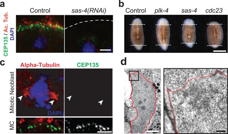
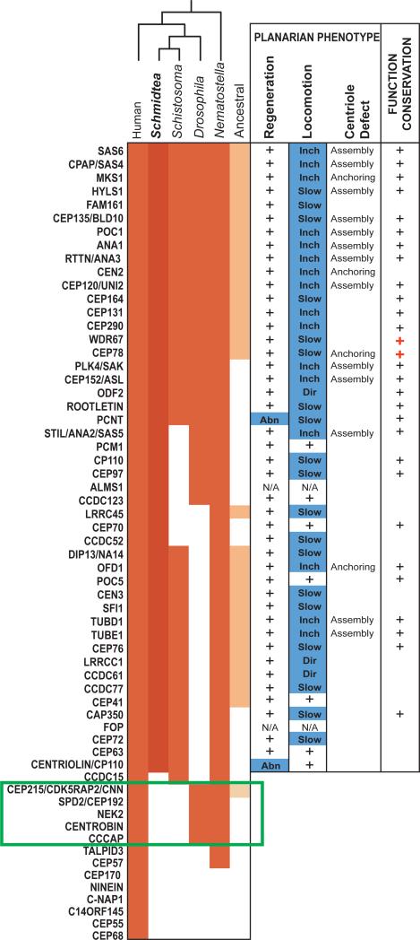
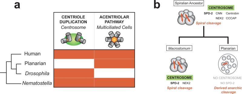
The centrosome, a cytoplasmic organelle formed by cylinder-shaped centrioles surrounded by a microtubule-organizing matrix, is a hallmark of animal cells. The centrosome is conserved and essential for the development of all animal species described so far. Here, we show that planarians, and possibly other flatworms, lack centrosomes. In planarians, centrioles are only assembled in terminally differentiating ciliated cells through the acentriolar pathway to trigger the assembly of cilia. We identified a large set of conserved proteins required for centriole assembly in animals and note centrosome protein families that are missing from the planarian genome. Our study uncovers the molecular architecture and evolution of the animal centrosome and emphasizes the plasticity of animal cell biology and development.


Figures

Figure 1
Figure 1
Centrioles in planarians are present in multiciliated cells but not proliferating cells. (a) Immunofluorescence staining of ventral epidermis in control or sas-4(RNAi) animals; centrioles (green, anti-SMED-CEP135), cilia (red, anti-acetylated tubulin), nuclei (blue, DAPI). (b) Regenerating trunk fragments. Dashed lines: boundary between newly formed and preexisting tissues. (c) Immunofluorescence staining of control mitotic neoblast; centrioles (green, anti-SMED-CEP135), mitotic spindle (red, anti-alpha-tubulin), chromosomes (blue, DAPI). Arrowheads: spindle poles. Lower panel: centrioles in a multiciliated cell (MC) from the same section. (d) TEM view of control mitotic neoblast (3,200x). Red line: plasma membrane. Right panel: high magnification view (15,000x) of boxed area. Bar is 5 μm in a, c and d-left panel, 1mm in b, and 0.5 μm in d-right panel.
Figure 2
Figure 2
Planarian homologs of human centrosome components are required for centriole assembly in multiciliated cells. Left: conservation of centrosome components in animals (red). Ancestral eukaryotic genes are in orange. Green box: centrosome signature genes lost in the planarian Schmidtea mediterranea and the parasite flatworm Schistosoma mansoni but present in Drosophila. Middle: regeneration and locomotion phenotypes in planarians (+: no defect; Abn: abnormal; Inch: inchworming; Dir: abnormal direction of locomotion). Right: +: genes implicated in centriole duplication or ciliogenesis in other systems (red +: this study; S.O.M.).
Figure 3
Figure 3
(a) Conservation of the two main pathways for centriole assembly in animals. Centrosome duplication and the acentriolar pathway are highlighted in red when present. (b) Model of the correlation between loss of the centrosome and loss of the spiral cleavage pattern.

References

    1. Calarco-Gillam PD, et al. Cell. 1983;35:621. - PubMed
    1. Bettencourt-Dias M, et al. Curr Biol. 2005;15:2199. - PubMed
    1. Dawe HR, et al. J Cell Sci. 2007;120:7. - PubMed
    1. Basto R, et al. Cell. 2006;125:1375. - PubMed
    1. Azimzadeh J, Marshall WF. Current biology : CB. 2010;20:R816. - PMC - PubMed

Publication types

Substances