. 2011 Dec 15;124(Pt 24):4253-66.
doi: 10.1242/jcs.091009. Epub 2012 Jan 5.

The induction of a nucleoplasmic reticulum by prelamin A accumulation requires CTP:phosphocholine cytidylyltransferase-α

Affiliations

The induction of a nucleoplasmic reticulum by prelamin A accumulation requires CTP:phosphocholine cytidylyltransferase-α

Chris N Goulbourne et al. J Cell Sci. .

Abstract

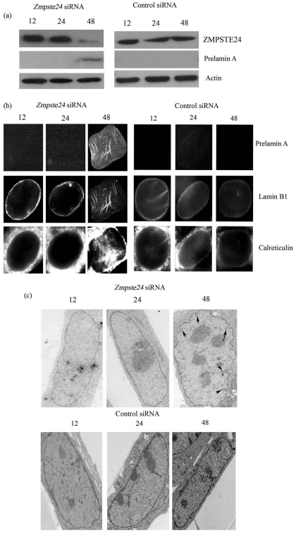
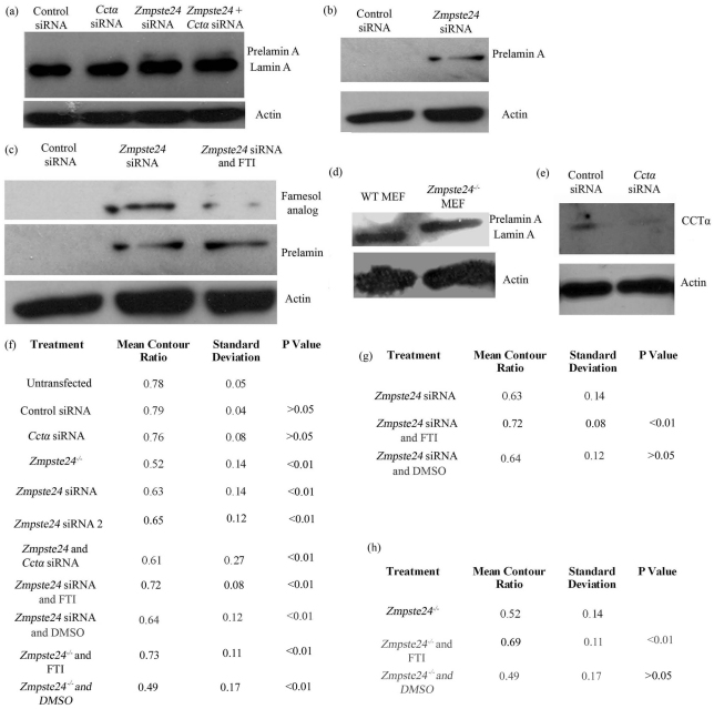
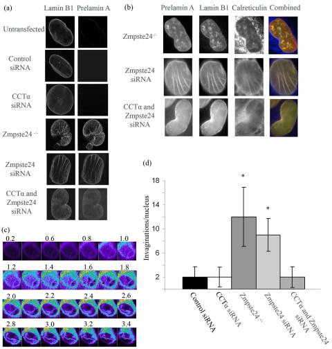
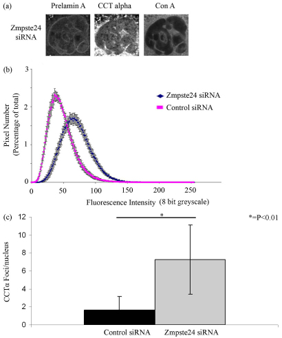
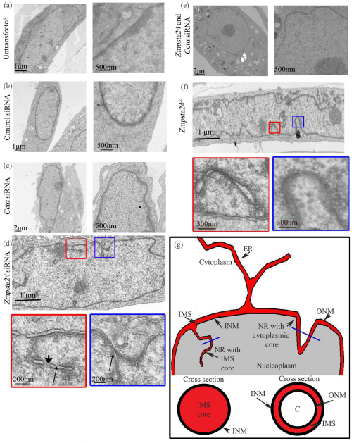
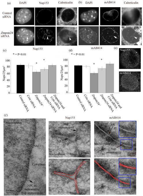
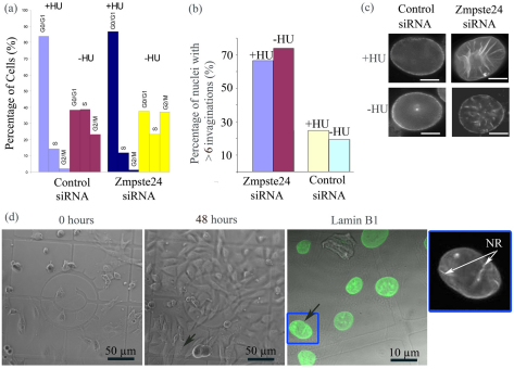
Farnesylated prelamin A accumulates when the final endoproteolytic maturation of the protein fails to occur and causes a dysmorphic nuclear phenotype; however, the morphology and mechanisms of biogenesis of these changes remain unclear. We show here that acute prelamin A accumulation after reduction in the activity of the ZMPSTE24 endoprotease by short interfering RNA knockdown, results in the generation of a complex nucleoplasmic reticulum that depends for its formation on the enzyme CTP:phosphocholine-cytidylyltransferase-α (CCT-α, also known as choline-phosphate cytidylyltransferase A). This structure can form during interphase, confirming that it is independent of mitosis and therefore not a consequence of disordered nuclear envelope assembly. Serial-section dual-axis electron tomography reveals that these invaginations can take two forms: one in which the inner nuclear membrane infolds alone with an inter membrane space interior, and the other in which an invagination of both nuclear membranes occurs, enclosing a cytoplasmic core. Both types of invagination can co-exist in one nucleus and both are frequently studded with nuclear pore complexes (NPC), which reduces NPC abundance on the nuclear surface.


Figures

Fig. 1.
Fig. 1.
siRNA knockdown of Zmpste24 at 48 hours leads to prelamin A accumulation and the development of NR. (a) Time course of Zmpste24 and control siRNA transfection examined at 12, 24 and 48 hours revealed that there was knockdown of ZMPSTE24 at 48 hours, which corresponded to the first detection of prelamin A. Control siRNA had no effect on ZMPSTE24 levels and no prelamin A was detected. (b) Immunofluorescence also revealed that the prelamin A was only detected at 48 hours in Zmpste24-siRNA-transfected cells and this also corresponded to the appearance of a convoluted nuclear boundary and the detection of NR development. The production of prelamin A and the development of an NR were absent in the control-siRNA-transfected cells. (c) TEM also revealed that it was only at 48 hours of Zmpste24 siRNA transfection that invaginations of the nuclear envelope could be seen to develop and there was the production of NR.
Fig. 2.
Fig. 2.
Accumulation of farnesylated prelamin A produces a dysmorphic nuclear phenotype that is rescued by FTI treatment. (a) Prelamin A accumulates after Zmpste24 siRNA transfection for 48 hours and Ccta siRNA transfection did not influence lamin levels. (b) Reactivity with a prelamin-A-specific antibody further supports that prelamin A accumulated after transfection. (c) Farnesyl analogue is detected in prelamin A in Zmpste24-siRNA-transfected cells and reduced in FTI-277-treated cells, indicating the prelamin A accumulating is farnesylated. (d) Lamin A/C antibody identifies just the higher band of prelamin A in Zmpste24−/− MEFs. (e) After 48 hours transfection with Ccta siRNA the level of CCT-α protein in the lysate has substantially decreased compared with control. (f) Dysmorphic shape (expressed as decreasing nuclear contour ratio) increases as prelamin A accumulates. The abnormality (lower contour ratio) is not rescued by co-transfection with Ccta siRNA. (g) FTI co-treatment significantly reduces the dysmorphic characteristic of ZmpSte24-siRNA-treated nuclei but does not rescue the CR completely to control level. (h) FTI co-treatment significantly reduces the dysmorphic characteristic of ZmpSte24−/− nuclei but does not rescue the CR completely to control level.
Fig. 3.
Fig. 3.
Immunofluorescence microscopy reveals the development of an NR in the context of prelamin A accumulation, which is dependent on CCT-α. (a) Prelamin A and lamin B1 colocalise around the nuclear envelope and at invaginations within the nucleoplasm and Ccta siRNA co-transfection ablates these intranuclear signals. (b) Prelamin A and lamin B1 colocalise with calreticulin in the nucleus and Ccta siRNA prevents the intranuclear NR signals, which support the presence of invaginations. (c) An example of a z-stack (numbers above images represent depth in micrometres) of nuclei labelled with calreticulin, used to quantify NR as illustrated in Fig. 3d. (d) NR was significantly (P<0.01) more abundant in prelamin A accumulating cells and co-transfection with Ccta siRNA reduced their frequency to control levels.
Fig. 4.
Fig. 4.
CCT-α foci develop along the prelamin-A-induced NR. (a) After Zmpste24 siRNA transfection CCT-α was found as foci along the invaginations that partly colocalised with prelamin A. (b) A frequency histogram of CCT-α intensity was calculated for each nucleus (ten nuclei per treatment). Zmpste24 siRNA transfection resulted in a shift to the right, indicating a greater abundance of higher intensity CCT-α pixels. (c) CCT-α foci were quantified and showed a significantly increased abundance after Zmpste24 siRNA transfection.
Fig. 5.
Fig. 5.
Morphology of the NR as revealed by TEM. (a) Untreated, (b) control siRNA and (c) Ccta-siRNA-transfected nuclei had an approximately circular nuclear membrane with few invaginations. (d) Zmpste24 siRNA transfection resulted in nuclei with invaginations (long thin arrows) that were perforated (short thick arrow). (e) Co-transfection with Zmpste24 and Ccta siRNA abolished NR development but the boundary of the nucleus was still convoluted. (f) Zmpste24−/− cells exhibited single-membrane nuclear invaginations (red box) and double-membrane invaginations with a cytoplasmic core (blue box). (g) Schematic illustrating the two types of invaginations that develop as part of the NR when farnesylated prelamin A accumulates.
Fig. 6.
Fig. 6.
Dual-axis electron tomography of type I NR reveals the presence of regular pores. (a) Dual-axis electron tomography of a 250 nm thick section revealed that the NR contained regularly sized pores, with similar dimensions to NPCs. (b) Serial section dual-axis electron tomography of seven 120 nm thick sections revealed that the NR was sheet like and contained many presumptive NPCs. Red is NR, purple is nuclear membrane and brown is cell membrane. (c) The connection to the nuclear membrane was revealed by an infolding of just the inner nuclear membrane (red represents NR and nuclear membrane in this case). CM, cell membrane; NM, nuclear membrane; NPC, nuclear pore complex; ONM, outer nuclear membrane; INM, inner nuclear membrane; IMS, inter-membrane space.
Fig. 7.
Fig. 7.
Nucleoporins colocalise with the NR. (a) Nup153immunofluorescence revealed a uniform and peripheral staining pattern around the nuclear membrane in control-siRNA-transfected cells. After Zmpste24 siRNA transfection more intranuclear nucleoporin signal could be seen. This signal colocalised with a gap in the DAPI signal and nucleoplasmic calreticulin (white arrowhead), indicating the presence of an invagination. (b) The same pattern was observed with the mAb414 that showed uniform peripheral staining during control siRNA transfection but contained intranuclear signals that colocalised with invaginations after Zmpste24 siRNA transfection. The frequency of Nup153 (c) and mAb414 (d) nucleoporin foci in a measured area of the nucleus was quantified. This approach revealed that prelamin A accumulation resulted in a significantly decreased abundance of nucleoporin foci on the nuclear surface and co-transfection of Zmpste24 and Ccta siRNA restored the foci for both nucleoporins on the nuclear surface. (e) Example of grazing sections of Nup153- and mAb414-stained nuclei as acquired in order to quantify the frequency of nucleoporins at the periphery; n=100 nuclei per treatment. (f) Immunogold EM of Nup153 and mAb414 antibody show regular peripheral staining at nuclear pore complexes. Nup153 and mAb414 gold particles associate with NR of cells transfected with Zmpste24 siRNA.
Fig. 8.
Fig. 8.
NR development is independent of mitosis. (a) Treatment with HU arrests cells in the G0–G1 phase of the cell cycle, preventing them passing through mitosis. (b,c) The treatment with HU had no significant effect on the number of invaginations. (d) Live-cell imaging of cells that had been transfected with Zmpste24 siRNA showing examples of cells that had not passed through mitosis but still developed an NR.

Similar articles

Cited by

References

    1. Bergo M. O., Gavino B., Ross J., Schmidt W. K., Hong C., Kendall L. V., Mohr A., Meta M., Genant H., Jiang Y., et al. (2002). Zmpste24 deficiency in mice causes spontaneous bone fractures, muscle weakness, and a prelamin A processing defect. Proc. Natl. Acad. Sci. USA 99, 13049-13054 - PMC - PubMed
    1. Bkaily G., Avedanian L., Jacques D. (2009). Nuclear membrane receptors and channels as targets for drug development in cardiovascular diseases. Can. J. Physiol. Pharmacol. 87, 108-119 - PubMed
    1. Bootman M. D., Fearnley C., Smyrnias I., MacDonald F., Roderick H. L. (2009). An update on nuclear calcium signalling. J. Cell Sci. 122, 2337-2350 - PubMed
    1. Busch A., Kiel T., Heupel W. M., Wehnert M., Hubner S. (2009). Nuclear protein import is reduced in cells expressing nuclear envelopathy-causing lamin A mutants. Exp. Cell Res. 315, 2373-2385 - PubMed
    1. Bussolati G., Marchio C., Gaetano L., Lupo R., Sapino A. (2008). Pleomorphism of the nuclear envelope in breast cancer: a new approach to an old problem. J. Cell Mol. Med. 12, 209-218 - PMC - PubMed

Publication types

MeSH terms