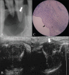
Ultrasonography with color Doppler and power Doppler in the diagnosis of periapical lesions
- PMID: 22223940
- PMCID: PMC3249943
- DOI: 10.4103/0971-3026.90688
Ultrasonography with color Doppler and power Doppler in the diagnosis of periapical lesions
Abstract
Aim: To evaluate the efficacy of ultrasonography (USG) with color Doppler and power Doppler applications over conventional radiography in the diagnosis of periapical lesions.
Materials and methods: Thirty patients having inflammatory periapical lesions of the maxillary or mandibular anterior teeth and requiring endodontic surgery were selected for inclusion in this study. All patients consented to participate in the study. We used conventional periapical radiographs as well as USG with color Doppler and power Doppler for the diagnosis of these lesions. Their diagnostic performances were compared against histopathologic examination. All data were compared and statistically analyzed.
Results: USG examination with color Doppler and power Doppler identified 29 (19 cysts and 10 granulomas) of 30 periapical lesions accurately, with a sensitivity of 100% for cysts and 90.91% for granulomas and a specificity of 90.91% for cysts and 100% for granulomas. In comparison, conventional intraoral radiography identified only 21 lesions (sensitivity of 78.9% for cysts and 45.4% for granulomas and specificity of 45.4% for cysts and 78.9% for granulomas). There was definite correlation between the echotexture of the lesions and the histopathological features except in one case.
Conclusions: USG imaging with color Doppler and power Doppler is superior to conventional intraoral radiographic methods for diagnosing the nature of periapical lesions in the anterior jaws. This study reveals the potential of USG examination in the study of other jaw lesions.
Keywords: Color Doppler; conventional intraoral radiography; histopathology; periapical lesions; power Doppler; ultrasound.
Conflict of interest statement
Figures


References
-
- Aggarwal V, Logani A, Shah N. The evaluation of computed tomography scans and ultrasounds in the differential diagnosis of periapical lesions. J Endod. 2008;34:1312–5. - PubMed
-
- Bender IB, Seltzer S. Roentgenographic and direct observation of experimental lesions in bone. J Am Dent Assoc. 1961;87:708–16.
-
- Bender IB, Seltzer S. Roentgenographic and direct observation of experimental lesions in bone. J Am Dent Assoc. 1961;87:708–16.
-
- Sumer AP, Danaci M, Ozen Sandikçi E, Sumer M, Celenk P. Ultrasonography and doppler ultrasonogrsaphy in the evaluation intraosseous lesions of the jaws. Dentomaxillofac Radiol. 2009;38:23–7. - PubMed
-
- Cotti E, Campisi G, Ambu R, Dettori C. Ultrasound real-time imaging in the differential diagnosis of periapical lesions. Int Endod J. 2003;36:556–63. - PubMed
LinkOut - more resources
Full Text Sources

