

Hepatogenic differentiation of mesenchymal stem cells induced by insulin like growth factor-I
- PMID: 22224170
- PMCID: PMC3251745
- DOI: 10.4252/wjsc.v3.i12.113
Hepatogenic differentiation of mesenchymal stem cells induced by insulin like growth factor-I
Abstract
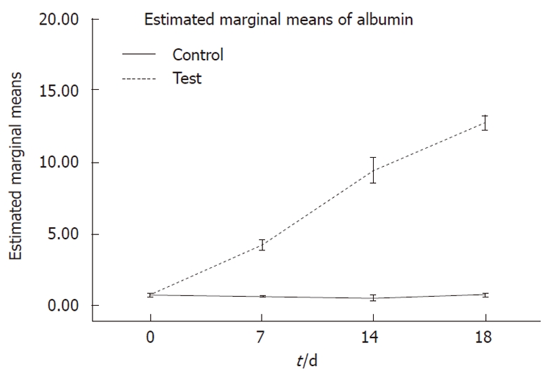
Aim: To improve hepatic differentiation of human mesenchymal stem cell (MSC) using insulin growth factor 1 (IGF-I), which has important role in liver development, hepatocyte differentiation and function.
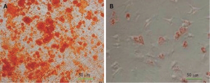
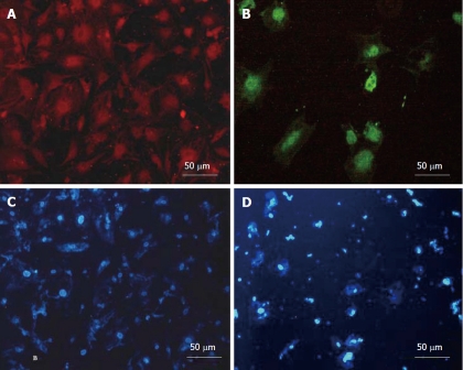
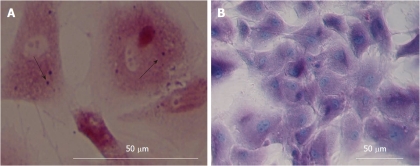
Methods: Bone marrow of healthy donors was aspirated from the iliac crest. The adherent cells expanded rapidly and were maintained with periodic passages until a relatively homogeneous population was established. The identification of these cells was carried out by immunophenotype analysis and differentiation potential into osteocytes and adipocytes. To effectively induce hepatic differentiation, we designed a protocol based on a combination of IGF-I and liver specific factors (hepatocyte growth factor, oncostatin M and dexamethasone). Morphological features, hepatic functions and cytological staining were assessed to evaluate transdifferentiation of human marrow-derived MSCs.
Results: Flow cytometric analysis and the differentiation potential into osteoblasts and adipocytes showed that more than 90% of human MSCs which were isolated and expanded were positive by specific markers and functional tests. Morphological assessment and evaluation of glycogen storage, albumin and α-feto protein expression, as well as albumin and urea secretion revealed a statistically significant difference between the experimental groups and control.
Conclusion: In vitro differentiated MSCs using IGF-I were able to display advanced liver metabolic functions, supporting the possibility of developing them as potential alternatives to primary hepatocytes.
Keywords: Differentiation; Hepatocyte; Human; Insulin-like growth factor 1; Mesenchymal stem cell.
Figures


Similar articles
-
Insulin-like growth factor 1 (IGF-I) improves hepatic differentiation of human bone marrow-derived mesenchymal stem cells.Cell Biol Int. 2011 Nov;35(11):1169-76. doi: 10.1042/CBI20110016. Cell Biol Int. 2011. PMID: 21910691
-
Useful properties of undifferentiated mesenchymal stromal cells and adipose tissue as the source in liver-regenerative therapy studied in an animal model of severe acute fulminant hepatitis.Cytotherapy. 2015 Aug;17(8):1052-65. doi: 10.1016/j.jcyt.2015.04.010. Cytotherapy. 2015. PMID: 26139545
-
In vitro hepatic differentiation of human bone marrow mesenchymal stem cells under differential exposure to liver-specific factors.Transl Res. 2009 Sep;154(3):122-32. doi: 10.1016/j.trsl.2009.05.007. Epub 2009 Jun 24. Transl Res. 2009. PMID: 19665688
-
The effect of dimethyl sulfoxide on hepatic differentiation of mesenchymal stem cells.Artif Cells Nanomed Biotechnol. 2016;44(1):157-64. doi: 10.3109/21691401.2014.928778. Epub 2014 Jun 30. Artif Cells Nanomed Biotechnol. 2016. PMID: 24978442
-
High-purity hepatic lineage differentiated from dental pulp stem cells in serum-free medium.J Endod. 2012 Apr;38(4):475-80. doi: 10.1016/j.joen.2011.12.011. Epub 2012 Jan 28. J Endod. 2012. PMID: 22414832
Cited by
-
The differentiation of human multipotent adult progenitor cells into hepatocyte-like cells induced by coculture with human hepatocyte line L02.Ann Surg Treat Res. 2015 Jan;88(1):1-7. doi: 10.4174/astr.2015.88.1.1. Epub 2014 Dec 26. Ann Surg Treat Res. 2015. PMID: 25553318 Free PMC article.
-
Therapeutic potential of mesenchymal stem cells for oral and systemic diseases.Dent Clin North Am. 2012 Jul;56(3):651-75. doi: 10.1016/j.cden.2012.05.006. Dent Clin North Am. 2012. PMID: 22835544 Free PMC article. Review.
-
Synergistic Hepatoprotective Effects of Mesenchymal Stem Cells and Platelet-Rich Plasma in a Rat Model of Bile Duct Ligation-Induced Liver Cirrhosis.Cells. 2024 Feb 26;13(5):404. doi: 10.3390/cells13050404. Cells. 2024. PMID: 38474368 Free PMC article.
-
Human Hepatocyte Transplantation: Three Decades of Clinical Experience and Future Perspective.Stem Cells Transl Med. 2024 Mar 15;13(3):204-218. doi: 10.1093/stcltm/szad084. Stem Cells Transl Med. 2024. PMID: 38103170 Free PMC article. Review.
-
Differentiation Potential of Breast Milk-Derived Mesenchymal Stem Cells into Hepatocyte-Like Cells.Tissue Eng Regen Med. 2017 Jul 27;14(5):587-593. doi: 10.1007/s13770-017-0066-x. eCollection 2017 Oct. Tissue Eng Regen Med. 2017. PMID: 30603512 Free PMC article.
References
-
- Dietreich D. Strategies for management of HCV/HIV coinfection. 3rd ed. PDR. 2005. pp. 101–113.
-
- Rozga J. Liver support technology--an update. Xenotransplantation. 2006;13:380–389. - PubMed
-
- Jalan R. Acute liver failure: current management and future prospects. J Hepatol. 2005;42 Suppl:S115–S123. - PubMed
-
- Nussler A, Konig S, Ott M, Sokal E, Christ B, Thasler W, Brulport M, Gabelein G, Schormann W, Schulze M, et al. Present status and perspectives of cell-based therapies for liver diseases. J Hepatol. 2006;45:144–159. - PubMed
-
- Selden C, Hodgson H. Cellular therapies for liver replacement. Transpl Immunol. 2004;12:273–288. - PubMed
LinkOut - more resources
Full Text Sources
