Review
. 2012:30:313-35.
doi: 10.1146/annurev-immunol-020711-075015. Epub 2012 Jan 6.

Reflex principles of immunological homeostasis

Affiliations
Review

Reflex principles of immunological homeostasis

Ulf Andersson et al. Annu Rev Immunol. 2012.

Abstract

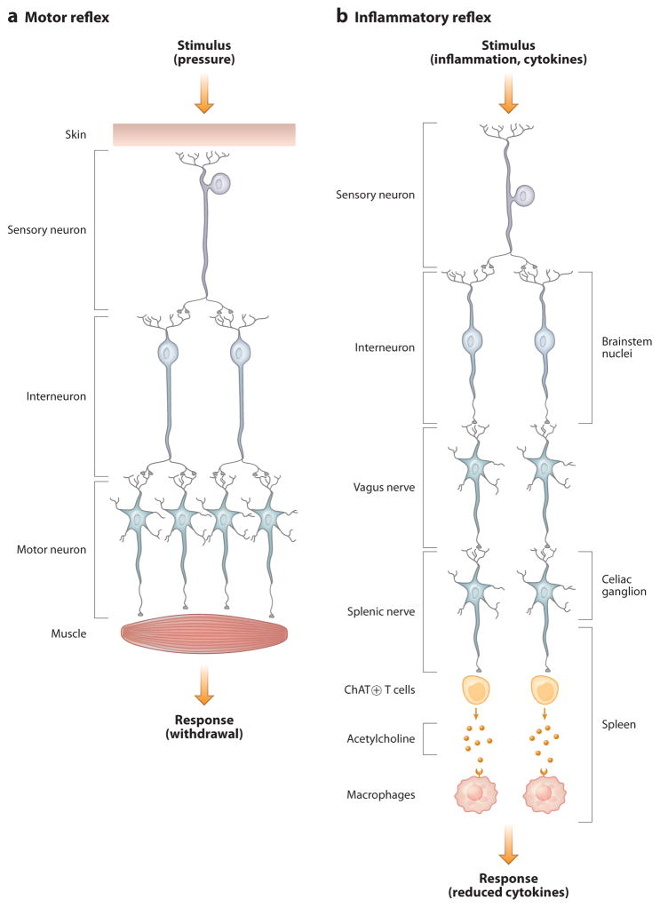
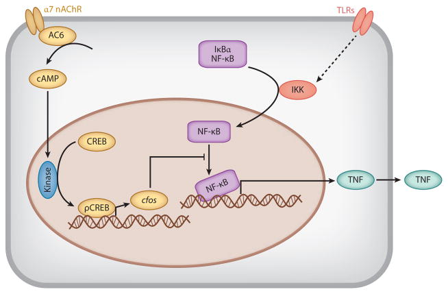
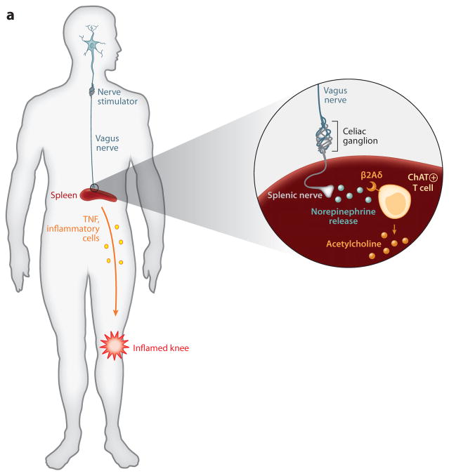
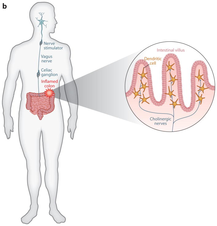
The reasoning that neural reflexes maintain homeostasis in other body organs, and that the immune system is innervated, prompted a search for neural circuits that regulate innate and adaptive immunity. This elucidated the inflammatory reflex, a prototypical reflex circuit that maintains immunological homeostasis. Molecular products of infection or injury activate sensory neurons traveling to the brainstem in the vagus nerve. The arrival of these incoming signals generates action potentials that travel from the brainstem to the spleen and other organs. This culminates in T cell release of acetylcholine, which interacts with α7 nicotinic acetylcholine receptors (α7 nAChR) on immunocompetent cells to inhibit cytokine release in macrophages. Herein is reviewed the neurophysiological basis of reflexes that provide stability to the immune system, the neural- and receptor-dependent mechanisms, and the potential opportunities for developing novel therapeutic devices and drugs that target neural pathways to treat inflammatory diseases.


Figures

Figure 1
Figure 1
Cellular basis of neural reflex circuits. (a) A simple motor reflex is composed of a sensory neuron that is activated by a stimulus (in this example, pressure). Sensory axons project to dendrites of interneurons, which in turn relay excitatory or inhibitory signals to corresponding muscle groups that produce withdrawal. (b) The inflammatory reflex is composed of sensory neurons traveling in the vagus nerve that are activated by products of infection (e.g., LPS) or inflammation (e.g., IL-1β). These project to brainstem nuclei, where interneurons relay the signals to the motor nuclei of the vagus nerve. The efferent signals travel in the vagus nerve to the celiac ganglion, where they interact with the cell bodies that give rise to axons that project in the splenic nerve. Action potentials lead to the release of norepinephrine, which activates acetylcholine release by a subset of T lymphocytes. Acetylcholine interacts with α7 nicotinic acetylcholine receptor (α7 nAChR) expressed in red pulp and marginal-zone macrophages to inhibit cytokine release.
Figure 2
Figure 2
Mechanism of inhibition of cytokine release mediated by α7 nicotinic acetylcholine receptor (α7 nAChR).
Figure 3
Figure 3
Neurons regulate innate immunity in Caenorhabditis elegans. (a) Wild-type C. elegans infected with bacterial pathogens die, but OCTR-1 knockout animals are significantly protected from lethal infection. (b) The mechanism depends on the role of OCTR-1-expressing neurons to inhibit innate gene expression required for the unfolded protein response.
Figure 4
Figure 4
Vagus nerve regulates HMGB1 release and lethality during sepsis. (a) Control mice subjected to lethal peritonitis secondary to a perforated cecum overexpress HMGB1, which mediates lethal organ injury. (b) Cutting the vagus, which normally maintains immunological homeostasis by transmitting signals that inhibit HMGB1 release, leads to significantly higher levels of HMGB1 (and other cytokines, not shown) and higher mortality rates. (c) Electrically stimulating the vagus nerve enhances the inhibitory signals and suppresses HMGB1, which improves survival. (d) The vagus nerve can also be stimulated pharmacologically using agents that target the cholinergic brain networks to increase vagus nerve activity. Vagus nerve activity is increased by CNI-1493, selective M1 receptor agonists, and centrally acting acetylcholinesterase inhibitors. These effectively increase the inhibitory vagus nerve signals that suppress HMGB1 levels and improve survival.
Figure 5
Figure 5
Implanted vagus nerve stimulators in arthritis and inflammatory bowel disease. (a) Vagus nerve–stimulating prototypes are being tested in the clinic to determine whether electrically stimulating the vagus nerve in humans will confer protection against cytokine-mediated inflammatory tissue damage. The approach is based on principles established in animal models that the vagus nerve regulates the activity of splenic neurons, which culminate in norepinephrine release in the spleen. Norepinephrine interacts with β2 adrenergic receptors expressed by T cells that, in turn, release acetylcholine, which inhibits cytokine release by signaling through α7 nAChR. At least two major effects of this neural mechanism prevent tissue injury: decreased release of TNF and other proinflammatory cytokines and decreased expression of adhesion molecules, HLA-DR, and other activation markers on monocytes as they transit the spleen en route to the joints or site of inflammation. (b) Vagus nerve stimulation in patients with inflammatory bowel disease may function through dual mechanisms, including the spleen pathway noted in panel a, and also by directly modulating inhibitory cholinergic neural signals to the intestine. Cytokine-producing cells that express α7 nAChR reside in the intestinal villi, where they lie in close proximity to acetylcholine-producing neurons that are regulated by vagus nerve signals.
Figure 5
Figure 5
Implanted vagus nerve stimulators in arthritis and inflammatory bowel disease. (a) Vagus nerve–stimulating prototypes are being tested in the clinic to determine whether electrically stimulating the vagus nerve in humans will confer protection against cytokine-mediated inflammatory tissue damage. The approach is based on principles established in animal models that the vagus nerve regulates the activity of splenic neurons, which culminate in norepinephrine release in the spleen. Norepinephrine interacts with β2 adrenergic receptors expressed by T cells that, in turn, release acetylcholine, which inhibits cytokine release by signaling through α7 nAChR. At least two major effects of this neural mechanism prevent tissue injury: decreased release of TNF and other proinflammatory cytokines and decreased expression of adhesion molecules, HLA-DR, and other activation markers on monocytes as they transit the spleen en route to the joints or site of inflammation. (b) Vagus nerve stimulation in patients with inflammatory bowel disease may function through dual mechanisms, including the spleen pathway noted in panel a, and also by directly modulating inhibitory cholinergic neural signals to the intestine. Cytokine-producing cells that express α7 nAChR reside in the intestinal villi, where they lie in close proximity to acetylcholine-producing neurons that are regulated by vagus nerve signals.

Similar articles

Cited by

References

    1. Nathan C, Ding A. Nonresolving inflammation. Cell. 2010;140:871–82. - PubMed
    1. Loewi O. Über homorale Übertragbarkeit der Herznervenwirkung. Pflügers Archiv. 1921;189:239–42.
    1. Dale HH, Dudley HW. The presence of histamine and acetylcholine in the spleen of the ox and the horse. J Physiol. 1929;68:97–123. - PMC - PubMed
    1. Sherrington C. The Integrative Action of the Nervous System. New Haven, CT: Yale Univ. Press; 1906.
    1. Tracey KJ. The inflammatory reflex. Nature. 2002;420:853–59. - PubMed

Publication types