Review
. 2012:30:175-202.
doi: 10.1146/annurev-immunol-030409-101320. Epub 2012 Jan 3.

The response to and repair of RAG-mediated DNA double-strand breaks

Affiliations
Review

The response to and repair of RAG-mediated DNA double-strand breaks

Beth A Helmink et al. Annu Rev Immunol. 2012.

Abstract

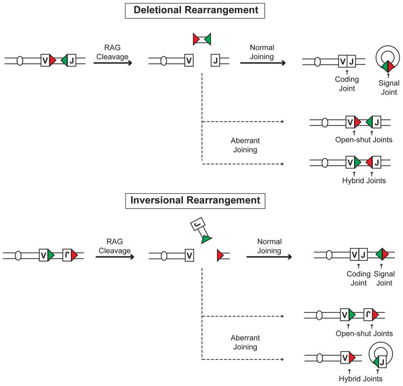
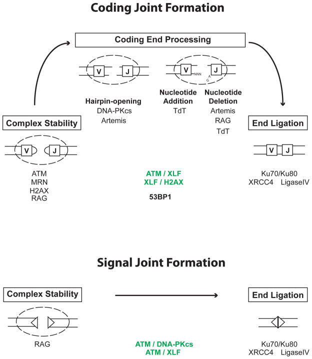
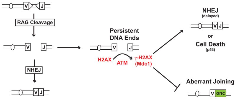
Developing lymphocytes must assemble antigen receptor genes encoding the B cell and T cell receptors. This process is executed by the V(D)J recombination reaction, which can be divided into DNA cleavage and DNA joining steps. The former is carried out by a lymphocyte-specific RAG endonuclease, which mediates DNA cleavage at two recombining gene segments and their flanking RAG recognition sequences. RAG cleavage generates four broken DNA ends that are repaired by nonhomologous end joining forming coding and signal joints. On rare occasions, these DNA ends may join aberrantly forming chromosomal lesions such as translocations, deletions and inversions that have the potential to cause cellular transformation and lymphoid tumors. We discuss the activation of DNA damage responses by RAG-induced DSBs focusing on the component pathways that promote their normal repair and guard against their aberrant resolution. Moreover, we discuss how this DNA damage response impacts processes important for lymphocyte development.


Figures

Figure 1
Figure 1. Types of rearrangements
Schematic of rearrangements that occur by deletion or inversion. RSs are indicated by colored triangles and the V and J gene segment by rectangles. The resulting normal signal and coding joints are depicted as are aberrant open-and-shut and hybrid joints.
Figure 2
Figure 2. Factors involved in signal and coding joint formation
RAG cleavage generates a pair of hairpin sealed coding ends (rectangles) and a pair of blunt phosphorylated signal ends (triangles). After RAG cleavage, coding ends and signal ends reside in complexes until they can be processed and joined. Coding end complexes are stabilized by ATM and MRN and may also be stabilized by H2AX and possibly RAG. ATM and MRN do not appear to be critical for signal end complex stability. However, ATM and DNA-PKcs have overlapping activities (shown in green) that are required for signal joint formation. Although the basis for this requirement is unknown, it could reflect the overlapping activity of ATM and DNA-PKcs in promoting signal end complex stability. RAG stays bound to signal ends in post-cleavage complexes generated in vitro and it has been suggested that RAG may stabilize signal ends in vivo, but a direct demonstration of this RAG function is lacking. Ku70/80 and DNA-PKcs, which are recruited early to DSBs, have been implicated in the stabilization of DNA ends prior to joining but their potential function in stabilizing RAG DSBs has not been examined. Hairpin sealed coding ends are opened through the endonuclease activity of Artemis. Its endonuclease activity requires association with and possibly phosphorylation by activated DNA-PKcs. Nucleotides can then be added to coding ends through the activity of TdT, and nucleotides can be lost through the nuclease activity of Artemis and possibly RAG and TdT. The joining of signal and coding ends is carried out by DNA Ligase IV and XRCC4 with Ku70/Ku80 potentially being important to align DNA ends prior to joining. Although Ku function is essential for both signal and coding joint formation, its specific activities at RAG DSBs have not been directly examined. Notably, ATM and XLF have overlapping activities (shown in green) that are also important for signal and coding joint formation. In addition, XLF has overlapping activities with H2AX that are required for coding joint formation. Whether these overlapping activities are also required for signal joint formation has not been examined. The nature of these overlapping activities of XLF and ATM or H2AX remains to be determined. 53BP1 is required for optimal coding joint formation for reasons that are not clear but may involve a function in long range joining and/or preventing DNA end resection.
Figure 3
Figure 3. Pathways that maintain genomic stability
RAG DSBs are repaired efficiently by NHEJ. However, rare un-repaired RAG DSB must eventually be resolved by NHEJ or, if they persist un-repaired, must promote cell death to prevent their aberrant resolution as potentially dangerous chromosomal lesions. In this regard, the ATM-dependent phosphorylation of H2AX, forming γ-H2AX, and MDC1, which binds to γ-H2AX, prevents persistent un-repaired RAG DSBs from being resected in a way that allows them to access aberrant joining pathways in G1-phase lymphocytes. Thus, H2AX may maintain the structure of the DNA end in G1-phase cells such that it is either joined normally by NHEJ or activates p53-mediated cell death if it persists unjoined.

References

    1. Fugmann SD, Lee AI, Shockett PE, Villey IJ, Schatz DG. The RAG proteins and V(D)J recombination: complexes, ends, and transposition. Annu Rev Immunol. 2000;18:495–527. - PubMed
    1. Oettinger MA. V(D)J recombination: on the cutting edge. Curr Opin Cell Biol. 1999;11:325–9. - PubMed
    1. Gellert M. V(D)J recombination: rag proteins, repair factors, and regulation. Annu Rev Biochem. 2002;71:101–32. - PubMed
    1. Desiderio S, Lin WC, Li Z. The cell cycle and V(D)J recombination. Curr Top Microbiol Immunol. 1996;217:45–59. - PubMed
    1. Rooney S, Chaudhuri J, Alt FW. The role of the non-homologous end-joining pathway in lymphocyte development. Immunol Rev. 2004;200:115–31. - PubMed

Publication types

Substances

LinkOut - more resources