. 2012 Jan 6:9:3.
doi: 10.1186/1742-4690-9-3.

Host cell species-specific effect of cyclosporine A on simian immunodeficiency virus replication

Affiliations

Host cell species-specific effect of cyclosporine A on simian immunodeficiency virus replication

Hiroaki Takeuchi et al. Retrovirology. .

Abstract

Background: An understanding of host cell factors that affect viral replication contributes to elucidation of the mechanism for determination of viral tropism. Cyclophilin A (CypA), a peptidyl-prolyl cis-trans isomerase (PPIase), is a host factor essential for efficient replication of human immunodeficiency virus type 1 (HIV-1) in human cells. However, the role of cyclophilins in simian immunodeficiency virus (SIV) replication has not been determined. In the present study, we examined the effect of cyclosporine A (CsA), a PPIase inhibitor, on SIV replication.

Results: SIV replication in human CEM-SS T cells was not inhibited but rather enhanced by treatment with CsA, which inhibited HIV-1 replication. CsA treatment of target human cells enhanced an early step of SIV replication. CypA overexpression enhanced the early phase of HIV-1 but not SIV replication, while CypA knock-down resulted in suppression of HIV-1 but not SIV replication in CEM-SS cells, partially explaining different sensitivities of HIV-1 and SIV replication to CsA treatment. In contrast, CsA treatment inhibited SIV replication in macaque T cells; CsA treatment of either virus producer or target cells resulted in suppression of SIV replication. SIV infection was enhanced by CypA overexpression in macaque target cells.

Conclusions: CsA treatment enhanced SIV replication in human T cells but abrogated SIV replication in macaque T cells, implying a host cell species-specific effect of CsA on SIV replication. Further analyses indicated a positive effect of CypA on SIV infection into macaque but not into human T cells. These results suggest possible contribution of CypA to the determination of SIV tropism.


Figures
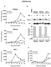
Figure 1
Figure 1
SIV and HIV-1 replication in human cells. (A) SIV and HIV-1 replication kinetics in CEM-SS cells. CEM-SS cells were infected with SIVagm (top panel), SIVmac (middle panel), and HIV-1 (bottom) in the absence (CsA[-], closed circles) or presence of 2.5 μM CsA (CsA[+], open circles). Virus production was monitored by measuring RT activity in the culture supernatants. (B) CypA incorporation into virions. Virus-containing supernatants were harvested from CsA-untreated and CsA-treated CEM-SS cells infected with SIVagm, SIVmac and HIV-1. A mock-infected sample was included as a control. CypA-specific band densities were quantified by densitometric scanning and are plotted in the lower panel. For each virus, the density of the band from CsA-untreated cells was defined as 100% and the ratio (%) of the density of the band from CsA-treated cells to that from CsA-untreated cells was calculated. The image of one representative blot is shown. (C) SIVagm replication kinetics in A3.01 cells.
Figure 2
Figure 2
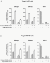
Effect of CsA treatment of target human T cells on SIV infection. (A) Effect of CsA treatment of producer human CEM-SS or target human LuSIV cells on viral infectivity. SIVagm, SIVmac, and HIV-1 from producer CEM-SS cells in the absence (Producer CEM-SS cell CsA [-]) or presence of CsA (Producer CEM-SS cell CsA [+]) were used to infect CsA-untreated (Target LuSIV cell CsA [-]) or CsA-treated target LuSIV cells (Target LuSIV cell CsA [+]). Luciferase activity in target LuSIV cells was measured 24 hr after infection. Relative infectivity is shown as the ratio (%) of the luciferase activity to that of viruses produced from CsA-untreated CEM-SS in CsA-untreated LuSIV cells. Mean values and standard deviations in four independent experiments are shown. (B) Effect of CsA treatment of producer or target human cells on viral replication. SIVagm, SIVmac, and HIV-1 from producer CEM-SS cells in the absence (Producer cell CsA [-]) or presence of CsA (Producer cell CsA [+]) were used to infect CsA-untreated (Target cell CsA [-]) or CsA-treated target CEM-SS cells (Target cell CsA [+]). Heat-inactivated virus was used as an infection control. Relative viral cDNA levels are shown as the ratio (%) of the viral cDNA levels to that of virus produced from CsA-untreated CEM-SS in CsA-untreated CEM-SS cells. Mean values and standard deviations in six independent experiments are shown.
Figure 3
Figure 3
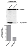
Immunoblot analysis of CypA expression. Lysates of CEM-SS (normal), CypA-KD cells were subjected to the analysis using anti-CypA antibodies. Anti-α-tubulin antibody was used as loading control. The ratio (%) of CypA band density in CypA-KD to that in normal CEM-SS is shown at the bottom panels. The image of one representative blot is shown.
Figure 4
Figure 4
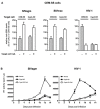
Effect of CypA knock-down on SIV and HIV-1 replication in human CEM-SS cells. (A) The amounts of viral cDNA synthesized after SIVagm (left panel), SIVmac239 (middle panel) or HIV-1 (right panel) infection. Viruses produced from CEM-SS cells were used to infect CsA-untreated or CsA-treated target CEM-SS, CypA-KD cells. Heat-inactivated virus was used as an infection control. The synthesized viral cDNA levels were measured by real-time PCR. Mean values and standard deviations in six independent experiments are shown. Relative viral cDNA levels are shown as the ratio (%) of the viral cDNA levels to that of viruses produced from CsA-untreated CEM-SS in CsA-untreated CEM-SS cells. (B) Replication of SIVagm (left panels) or HIV-1 (right panels) in CypA-KD CEM-SS cells. Viral production in normal CsA-untreated CEM-SS (closed circles), CsA-untreated (closed squares) or CsA-treated CypA-KD (open squares) was monitored by measuring RT activity in the culture supernatants.
Figure 5
Figure 5
Effect of CypB knock-down on HIV-1 and SIV replication in human CEM-SS T cells. (A) Immunoblot analysis of CypB expression. Lysates of CEM-SS (normal), CypA-KD, and CypB-KD cells were subjected to the immunoblot analysis using anti-α-tubulin, anti-CypA and anti-CypB antibodies (Abcam Inc., Cambridge, MA) (left panel). CypA- and CypB-specific band densities were quantified by densitometric scanning and are plotted in the right panels. The image of one representative blot is shown. (B) Replication of SIVagm (left panels) or HIV-1 (right panels) in CypB-KD CEM-SS cells. Viral production in normal CEM-SS (closed circles) or CypB-KD cells (closed triangles) was monitored by measuring RT activity in the culture supernatants.
Figure 6
Figure 6
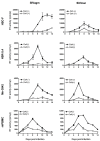
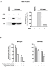
SIV replication in macaque cells. Cynomolgus macaque HSC-F, rhesus macaque HSR-5.4, pig-tailed macaque Mn-3942, and rhesus macaque PBMCs (rhPBMC) were infected with SIVagm (left panels) and SIVmac (right panels) and cultured in the absence (CsA[-], closed circles) or presence of 2.5 μM CsA (CsA[+], open circles). Virus production was monitored by measuring RT activity in the culture supernatants.
Figure 7
Figure 7
Effect of CsA treatment of macaque T cells on SIV infection. (A) Efficiency of CypA incorporation into virions from producer macaque HSC-F cells. Culture supernatants were harvested from CsA-untreated mock, and CsA-untreated and CsA-treated HSC-F cells infected with SIVagm. The CypA incorporation efficiency (right panel) is shown as described in the legend for Figure 1B. The image of one representative blot is shown. (B) Effect of CsA treatment of producer or target macaque HSC-F cells on SIV replication. SIVagm and SIVmac produced from HSC-F cells in the absence (Producer cell CsA [-]) or presence of CsA (Producer cell CsA [+]) was used to infect CsA-untreated (Target cell CsA [-]) or CsA-treated target HSC-F cells (Target cell CsA [+]). Heat-inactivated virus was used as an infection control. Relative viral cDNA levels are shown as the ratio (%) of the viral cDNA levels to that of virus produced from CsA-untreated HSC-F in CsA-untreated HSC-F cells. The synthesized viral cDNA levels were measured by real-time PCR. Mean values and standard deviations in four independent experiments are shown.
Figure 8
Figure 8
Effect of exogenous CypA expression on SIV and HIV-1 replication. (A) The amounts of viral cDNA synthesized after SIVagm (left panel) or HIV-1 (right panel) infection in human CEM-SS cells. Viruses produced from normal CEM-SS cells were used to infect CsA-untreated or CsA-treated target normal CEM-SS cells. Heat-inactivated virus was used as an infection control. Relative viral cDNA levels are shown as the ratio (%) of the viral cDNA levels to that of viruses produced from CsA-untreated CEM-SS in CsA-untreated CEM-SS cells. Mean values and standard deviations in three independent experiments are shown. (B) The amounts of viral cDNA after SIVagm infection in macaque HSC-F cells. SIVagm produced from normal HSC-F cells was used to infect CsA-untreated or CsA-treated target normal HSC-F cells. Heat-inactivated virus was used as an infection control. Relative viral cDNA levels are shown as the ratio (%) of the viral cDNA levels to that of viruses produced from CsA-untreated HSC-F in CsA-untreated HSC-F cells. Mean values and standard deviations in three independent experiments are shown.
Figure 9
Figure 9
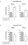
Infection of human and macaque cells with macaque- and human-derived SIV. (A) SIVagm and SIVmac produced from HSC-F cells in the absence or presence of CsA were used to infect CsA-untreated or CsA-treated target CEM-SS cells. Relative viral cDNA levels are shown as the ratio (%) of the viral cDNA levels to that of viruses produced from CsA-untreated HSC-F in CsA-untreated CEM-SS cells. Mean values and standard deviations in three independent experiments are shown. (B) SIVagm and SIVmac produced from CEM-SS cells in the absence or presence of CsA were used to infect CsA-untreated or CsA-treated target HSC-F cells. Relative viral cDNA levels are shown as the ratio (%) of the viral cDNA levels to that of viruses produced from CsA-untreated CEM-SS in CsA-untreated HSC-F cells. The relative viral cDNA levels synthesized were measured by real-time PCR. Mean values and standard deviations in three independent experiments are shown.

Similar articles

Cited by

References

    1. Gautsch JW, Elder JH, Schindler J, Jensen FC, Lerner RA. Structural markers on core protein p30 of murine leukemia virus: functional correlation with Fv-1 tropism. Proc Natl Acad Sci USA. 1978;75:4170–4174. doi: 10.1073/pnas.75.9.4170. - DOI - PMC - PubMed
    1. Rein A, Kashmiri SV, Bassin RH, Gerwin BL, Duran-Troise G. Phenotypic mixing between N- and B-tropic murine leukemia viruses: infectious particles with dual sensitivity to Fv-1 restriction. Cell. 1976;7:373–379. doi: 10.1016/0092-8674(76)90166-5. - DOI - PubMed
    1. Towers G, Bock M, Martin S, Takeuchi Y, Stoye JP, Danos O. A conserved mechanism of retrovirus restriction in mammals. Proc Natl Acad Sci USA. 2000;97:12295–12299. doi: 10.1073/pnas.200286297. - DOI - PMC - PubMed
    1. Stremlau M, Owens CM, Perron MJ, Kiessling M, Autissier P, Sodroski J. The cytoplasmic body component TRIM5alpha restricts HIV-1 infection in Old World monkeys. Nature. 2004;427:848–853. doi: 10.1038/nature02343. - DOI - PubMed
    1. Keckesova Z, Ylinen LM, Towers GJ. The human and African green monkey TRIM5alpha genes encode Ref1 and Lv1 retroviral restriction factor activities. Proc Natl Acad Sci USA. 2004;101:10780–10785. doi: 10.1073/pnas.0402474101. - DOI - PMC - PubMed

Publication types

LinkOut - more resources