. 2012 Jan 20;148(1-2):285-95.
doi: 10.1016/j.cell.2011.12.007. Epub 2012 Jan 5.

Generation of chimeric rhesus monkeys

Affiliations

Generation of chimeric rhesus monkeys

Masahito Tachibana et al. Cell. .

Abstract

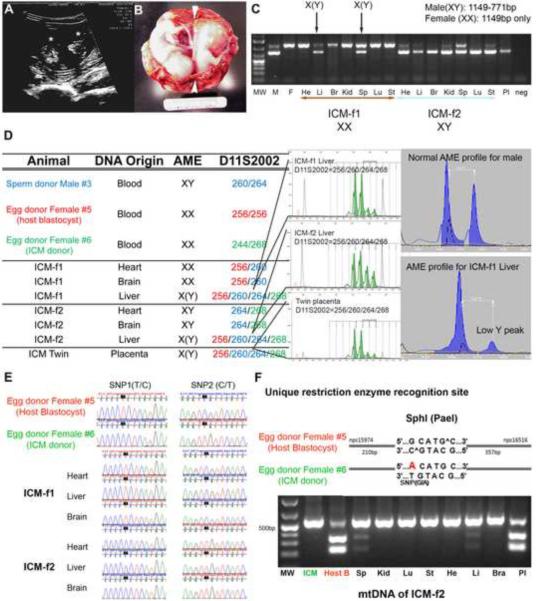
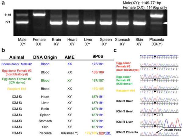
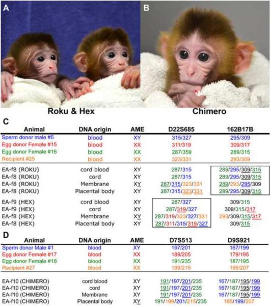
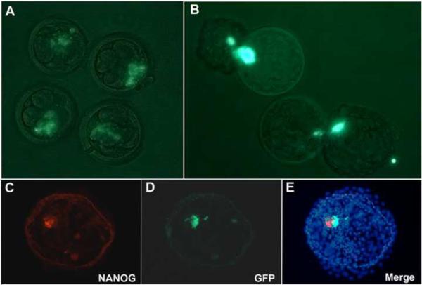
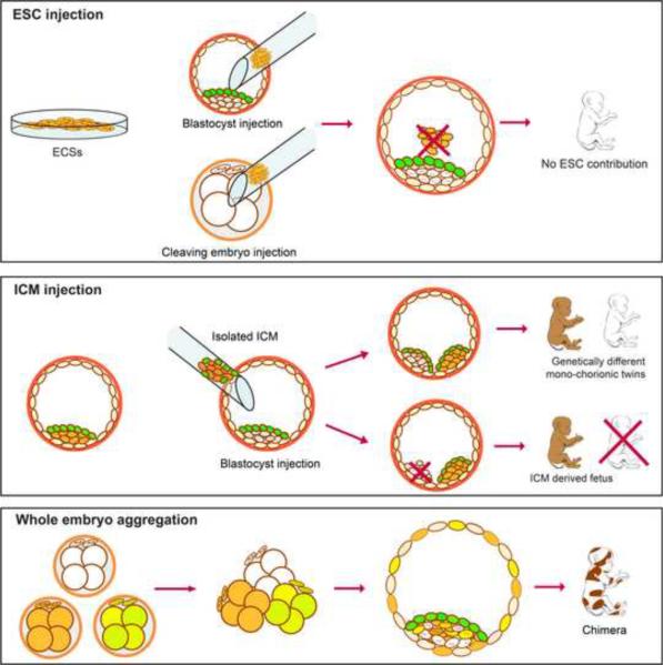
Totipotent cells in early embryos are progenitors of all stem cells and are capable of developing into a whole organism, including extraembryonic tissues such as placenta. Pluripotent cells in the inner cell mass (ICM) are the descendants of totipotent cells and can differentiate into any cell type of a body except extraembryonic tissues. The ability to contribute to chimeric animals upon reintroduction into host embryos is the key feature of murine totipotent and pluripotent cells. Here, we demonstrate that rhesus monkey embryonic stem cells (ESCs) and isolated ICMs fail to incorporate into host embryos and develop into chimeras. However, chimeric offspring were produced following aggregation of totipotent cells of the four-cell embryos. These results provide insights into the species-specific nature of primate embryos and suggest that a chimera assay using pluripotent cells may not be feasible.


Figures

Figure 1
Figure 1
Mono-chorionic twin fetuses produced by injection of an ICM into a blastocyst A, Ultrasonography image of a twin pregnancy at 30 days of gestation. Asterisks depict individual fetuses. B, Morphological analysis of fetuses recovered on day 51 of gestation. Note that while two fetuses share single placenta, a thin septum (arrowheads) separates each fetal cavity indicating mono-chorionic but di-amniotic pregnancy. C, PCR amplification of ZFX and ZFY regions produced two DNA fragments (male and female). Detection of 771bp fragment in liver and spleen samples of ICM-f1 indicates presence of male cells in female organs. D, Analysis of D11S2002 and AME microsatellite loci detected the presence of 3 different alleles in livers and a placenta of fetuses. E, Chromatogram of the rhesus mtDNA DHV1 region showing informative SNPs. Fetus ICM-f1 originated from a host blastocyst while ICM-f2 developed from the injected ICM. F, mtDNA RFLP analysis. G allele in mtDNA of the host blastocyst egg donor female #5 is recognized and digested by SphI (PaeI) while an A allele in the injected ICM egg donor female #6 precludes restriction. MtDNA haplotype of egg donor female #5 was detected in the liver and spleen of the ICM-f2 fetus derived from the ICM egg donor female #6. Abbreviations in Fig. 1C: MW, M, F, He, Li, Br, Kid, Sp, Lu, St, Pl and neg indicate Molecular Weght, Male, Female. Heart, Liver, Brain, Kidney, Spleen, Lung, Stomach, Placenta and negative control, respectively. (See also Table S2 for detailed STR data)
Figure 2
Figure 2
Parentage analysis of offspring derived from ICM A. Gender-specific PCR analysis demonstrating normal profiles for rhesus male (2 fragments of 1149bp and 771pb size) and female (one 1149bp fragment) DNA samples. Analysis of tissues and organs from ICM-f3 fetus produced by ICM injection demonstrated that the fetus is male while the placenta is female. In addition, the placenta showed a faint 771bp fragment (arrowhead) indicating presence of male cells at low levels. B. Microsatellite genotyping within the STR locus 9P06 clearly demonstrating that the fetus originated from the injected ICM whereas placenta was mainly from the host blastocyst. AME STR locus showed a limited presence of male cells in the female placenta possibly indicating amniotic contribution from the transplanted ICM. C. Chromatogram of mtDNA DHV1 region demonstrating informative SNPs that can distinguish mitochondrial contribution in tissues. An mtDNA profile of the ICM-f3 fetus matched to the transplanted ICM, while the placental mitochondrial genome was mixture of the host blastocyst and ICM-f3. Limited mtDNA contribution from the fetus was also evident in the placental tissues. (See also Table S2 for detailed STR data)
Figure 3
Figure 3
Chimeric infants generated by whole embryo aggregation A and B, Live chimeric offspring (Roku, Hex indicating 6 in Japanese and Greek, and Chimero) each produced by aggregating of six individual embryos. The pictures were taken at 7 days after birth. C and D, Genetic analysis of blood and extraembryonic tissues based on microsatellite examination demonstrating presence of more than two alleles for each locus. (See also Tables S4, S6, and Figure S6)
Figure 4
Figure 4
Detection of ESCs in blastocysts developed from 4-cell embryos injected with GFP positive ESCs A, GFP expressing ESCs were injected into 4-cell embryos and placed between blastomeres. B, Blastocysts with GFP embedded cells. C–E, Immune staining for NANOG demonstrated that GFP expressing ESCs within an ICM were NANOG negative indicating premature differentiation. Original magnifications: A – E; ×200
Figure 5
Figure 5
Summary of chimera studies with monkey embryos and embryonic cells. Rhesus monkey ESCs as well as isolated ICMs, blastomeres or whole embryos were tested for their ability to incorporate into host embryos and generate chimeric offspring. Established ESCs and freshly isolated ICMs failed to produce chimeras when injected into host blastocysts. However, ICMs developed into separate fetuses with placental support from the host embryo. Aggregating of several 4-cell embryos efficiently produced live chimeric offspring. (See also Figure S7 and Movie S1)

Comment in

References

    1. Beddington RS, Robertson EJ. An assessment of the developmental potential of embryonic stem cells in the midgestation mouse embryo. Development. 1989;105:733–737. - PubMed
    1. Bradley A, Evans M, Kaufman MH, Robertson E. Formation of germ-line chimaeras from embryo-derived teratocarcinoma cell lines. Nature. 1984;309:255–256. - PubMed
    1. Brem G, Tenhumberg H, Krausslich H. Chimerism in cattle through microsurgical aggregation of morulae. Theriogenology. 1984;22:609–613. - PubMed
    1. Brons IG, Smithers LE, Trotter MW, Rugg-Gunn P, Sun B, Chuva de Sousa Lopes SM, Howlett SK, Clarkson A, Ahrlund-Richter L, Pedersen RA, et al. Derivation of pluripotent epiblast stem cells from mammalian embryos. Nature. 2007;448:191–195. - PubMed
    1. Byrne JA, Pedersen DA, Clepper LL, Nelson M, Sanger WG, Gokhale S, Wolf DP, Mitalipov SM. Producing primate embryonic stem cells by somatic cell nuclear transfer. Nature. 2007;450:497–502. - PubMed

Publication types

LinkOut - more resources