

Prognostic value of proliferation assay in the luminal, HER2-positive, and triple-negative biologic classes of breast cancer
- PMID: 22225836
- PMCID: PMC3496118
- DOI: 10.1186/bcr3084
Prognostic value of proliferation assay in the luminal, HER2-positive, and triple-negative biologic classes of breast cancer
Abstract
Introduction: Although the prognostic significance of proliferation in early invasive breast cancer has been recognized for a long time, recent gene-expression profiling studies have reemphasized its biologic and prognostic value and the potential application of its assessment in routine practice, particularly to define prognostic subgroups of luminal/hormone receptor-positive (HR+) tumors. This study aimed to assess the prognostic value of a proliferation assay by using Ki-67 immunohistochemistry as compared with mitotic count scores.
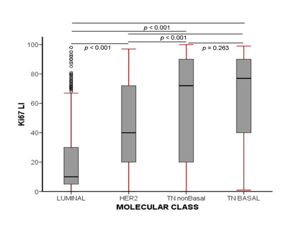
Method: Proliferation was assessed by using Ki-67 labeling index (Ki-67LI) and mitotic scores in a large (n = 1,550) and well-characterized series of clinically annotated primary operable invasive breast cancer with long-term follow-up. Tumors were phenotyped based on their IHC profiles into luminal/HR+, HER2+, and triple-negative (TN) classes. We used a split-sample development and validation approach to determine the optimal Ki-67LI cut-offs.
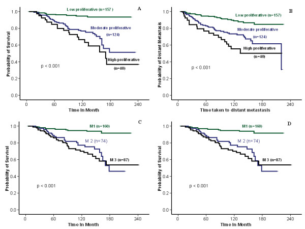
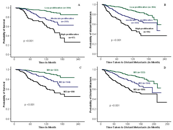
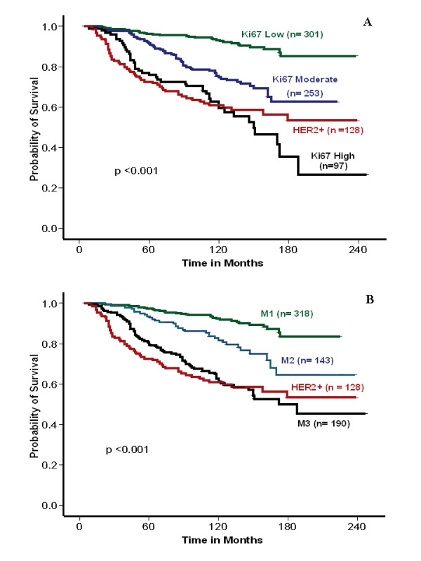
Results: The optimal cut-points of Ki-67LI were 10% and 50% for the luminal class. Both Ki7LI and MS were able to split luminal tumors into subgroups with significantly variable outcomes, independent of other variables. Neither mitotic count scores nor Ki-67LI was associated with outcome in the HER2+ or the TN classes.
Conclusions: Assessment of proliferation by using Ki-67LI and MS can distinguish subgroups of patients within luminal/hormone receptor-positive breast cancer significantly different in clinical outcomes. Overall, both Ki-67 LI and mitotic-count scores showed comparable results. The method described could provide a cost-effective method for prognostic subclassification of luminal/hormone receptor-positive breast cancer in routine clinical practice.
Figures
References
-
- Daidone MG, Silvestrini R. Prognostic and predictive role of proliferation indices in adjuvant therapy of breast cancer. J Natl Cancer Inst Monogr. 2001;30:27–35. - PubMed
-
- Wirapati P, Sotiriou C, Kunkel S, Farmer P, Pradervand S, Haibe-Kains B, Desmedt C, Ignatiadis M, Sengstag T, Schütz F, Goldstein DR, Piccart M, Delorenzi M. Meta-analysis of gene expression profiles in breast cancer: toward a unified understanding of breast cancer subtyping and prognosis signatures. Breast Cancer Res. 2008;10:R65. doi: 10.1186/bcr2124. - DOI - PMC - PubMed
-
- Weigelt B, Baehner FL, Reis-Filho JS. The contribution of gene expression profiling to breast cancer classification, prognostication and prediction: a retrospective of the last decade. J Pathol. 2010;220:263–80. - PubMed
-
- Paik S, Shak S, Tang G, Kim C, Baker J, Cronin M, Baehner FL, Walker MG, Watson D, Park T, Hiller W, Fisher ER, Wickerham DL, Bryant J, Wolmark N. A multigene assay to predict recurrence of tamoxifen-treated, node-negative breast cancer. N Engl J Med. 2004;351:2817–26. doi: 10.1056/NEJMoa041588. - DOI - PubMed
-
- Sotiriou C, Wirapati P, Loi S, Harris A, Fox S, Smeds J, Nordgren H, Farmer P, Praz V, Haibe-Kains B, Desmedt C, Larsimont D, Cardoso F, Peterse H, Nuyten D, Buyse M, Van de Vijver MJ, Bergh J, Piccart M, Delorenzi M. Gene expression profiling in breast cancer: understanding the molecular basis of histologic grade to improve prognosis. J Natl Cancer Inst. 2006;98:262–72. doi: 10.1093/jnci/djj052. - DOI - PubMed
Publication types
MeSH terms
Substances
Grants and funding
LinkOut - more resources
Full Text Sources
Medical
Research Materials
Miscellaneous
