. 2012 Jan 6:7:1.
doi: 10.1186/1749-8104-7-1.

Neurogenesis in the central olfactory pathway of adult decapod crustaceans: development of the neurogenic niche in the brains of procambarid crayfish

Affiliations

Neurogenesis in the central olfactory pathway of adult decapod crustaceans: development of the neurogenic niche in the brains of procambarid crayfish

Silvia Sintoni et al. Neural Dev. .

Abstract

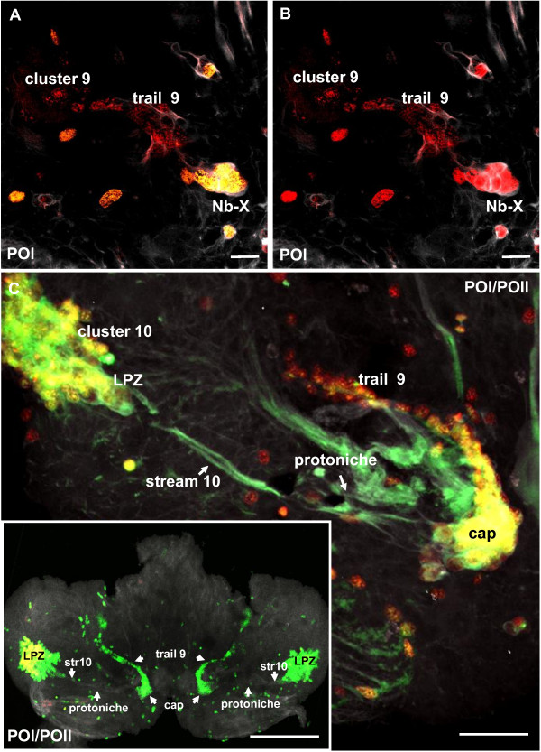
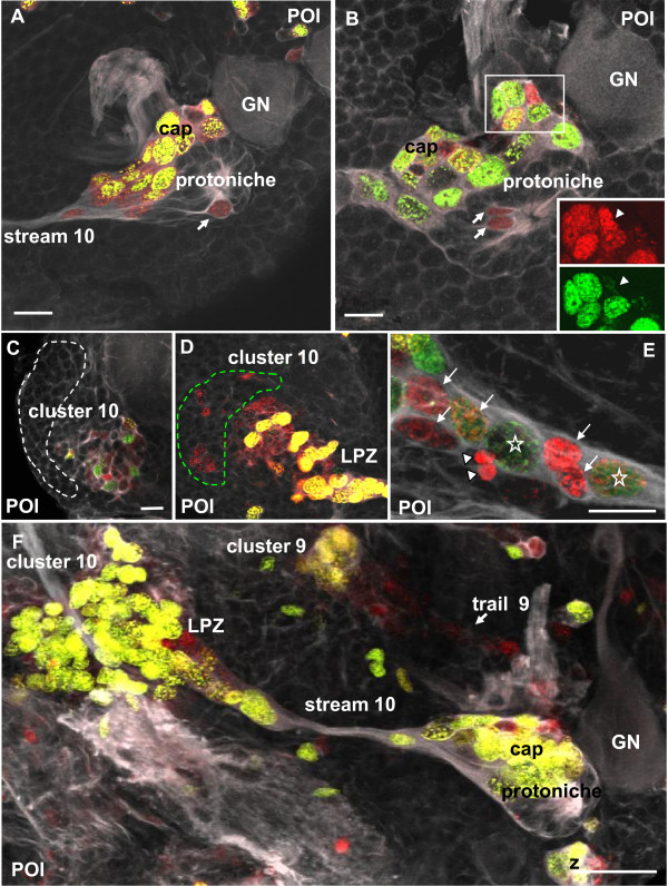
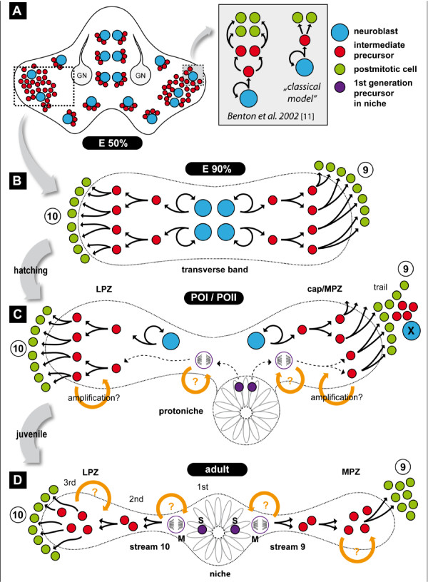
Background: In the decapod crustacean brain, neurogenesis persists throughout the animal's life. After embryogenesis, the central olfactory pathway integrates newborn olfactory local and projection interneurons that replace old neurons or expand the existing population. In crayfish, these neurons are the descendants of precursor cells residing in a neurogenic niche. In this paper, the development of the niche was documented by monitoring proliferating cells with S-phase-specific markers combined with immunohistochemical, dye-injection and pulse-chase experiments.

Results: Between the end of embryogenesis and throughout the first post-embryonic stage (POI), a defined transverse band of mitotically active cells (which we will term 'the deutocerebral proliferative system' (DPS) appears. Just prior to hatching and in parallel with the formation of the DPS, the anlagen of the niche appears, closely associated with the vasculature. When the hatchling molts to the second post-embryonic stage (POII), the DPS differentiates into the lateral (LPZ) and medial (MPZ) proliferative zones. The LPZ and MPZ are characterized by a high number of mitotically active cells from the beginning of post-embryonic life; in contrast, the developing niche contains only very few dividing cells, a characteristic that persists in the adult organism.

Conclusions: Our data suggest that the LPZ and MPZ are largely responsible for the production of new neurons in the early post-embryonic stages, and that the neurogenic niche in the beginning plays a subordinate role. However, as the neuroblasts in the proliferation zones disappear during early post-embryonic life, the neuronal precursors in the niche gradually become the dominant and only mechanism for the generation of new neurons in the adult brain.


Figures

Figure 1
Figure 1
The neurogenic system in the brain of the adult crayfish Procambarus clarkii (modified from [31]). Schematic diagram of the crayfish neurogenic system in the deutocerebrum. Neuronal precursors (first generation) with glial characteristics reside within the neurogenic niche where they divide symmetrically. Their daughters (second-generation precursors) migrate along tracts formed by the fibers of the niche cells, towards either the LPZ or the MPZ. At least one more division occurs in the LPZ and MPZ before the progeny (third and successive generations) differentiate into neurons. Abbreviations: LPZ, lateral proliferative zone; MPZ, medial proliferative zone.
Figure 2
Figure 2
Neurogenesis during early and mid-embryogenesis: BrdU labeling in whole mount embryos. (A, B) In earlier stages (about E38%), BrdU+ cells are homogeneously distributed throughout the germ band with pronounced accumulations in the appendage anlagen. The paired deutocerebral regions show an aggregation of labeled cells (dotted line). (C, D) During mid-embryogenesis (E50%), the embryos are characterized by a persistently high rate of mitotic activity. The deutocerebrum (DC, dotted line in (D)) stands out from the protocerebrum and contains large spherical BrdU+ cells. (E-G) Higher magnification of brain neuroblasts (NBs) and associated progeny, the ganglion mother cells (GMCs). Neuroblasts can be found in early prophase (F) or in early telophase (G). Abbreviations: A1, anlage of antenna 1; A2, anlage of antenna 2; OA, optic anlagen; PC, protocerebrum. Scale bars: 50 μm (A-D), 10 μm (E-G).
Figure 3
Figure 3
Neurogenesis during late embryogenesis: BrdU labeling in whole mount brains. (A) Arrangement of BrdU+ cells in the brain and optic anlagen of an E70% embryo. In the optic lobe anlagen (OL; and green triangles), in the protocerebrum (orange triangles) and in the deutocerebrum (white triangles) BrdU+ cells show a bilaterally symmetrical arrangement. (B) Higher magnification of neuroblasts (asterisks) in the medial protocerebrum (MPC) and the lateral protocerebrum (LPC; optic lobe anlagen). Scale bar: 60 μm (A), 15 μm (B).
Figure 4
Figure 4
Hatching and postembryonic stages, the emergence of the protoniche. (A-F) S-phase labeling with EdU (green), and immunohistochemistry for tyrosinated tubulin (white; confocal laser-scan images). (A) Overview of the brain during hatching/POI showing the transverse deutocerebral band of mitotic cells (encircled). (B) Higher magnification of the transverse band and the forming protoniche close to the giant neuron (GN) in the left side of the brain. (C-E) Two additional specimens showing the transverse band of mitotic cells and the protoniche in the left side of the brain. Panels (D, F) show the green channels only (EdU) of (C, E), respectively. In contrast to the numbers of dividing cells in the cap, the protoniche contains fewer and smaller EdU+ cells ( arrowheads in B, C, E). (G, H) Immunostaining for glutamine synthetase (in white), a glia cell marker, shows how the main structures of the neurogenic system (the protoniche and cap) all display immunolabeling, from the beginning of the post-embryonic life (G) to the POII stage (H) (both panels show the left side of the brain). Abbreviations: cl 10, cell cluster 10; GN, medial giant neuron. Scale bars: 100 μm (A), 20 μm (B-H).
Figure 5
Figure 5
The emergence of the niche core during hatching, immunolocalization of tyrosinated tubulin and the nuclear stain YOYO (magenta). (A) Ventral view of the brain showing two forming protoniches that are located bilaterally symmetrical on the surface of the deutocerebrum (square on the right, circle on the left) close to the medial giant neuron (GN). In the brain, clusters composed of neuroblasts and their progeny are still patchily dispersed (white stars). Arrowheads identify nerve roots. (B) Higher magnification of the protoniche boxed in (A). The arrow points to microtubules that converge in the center of the protoniche. (C) Another example of a protoniche located close to the giant neuron (GN). (D) Deeper focus of the specimen in (C) showing a cell in M-phase (arrow). (E) Within the protoniche, cells that surround the central fibrous area have very condensed, small nuclei (arrows). The cells in the cap are spherical but vary greatly in size. (F) Dividing cells in the protoniche close to surrounding the central fibrous area (arrows). Scale bars: 20 μm (A, B, E), 10 μm (C, D, F).
Figure 6
Figure 6
The protoniche and cap in the POI stage, left side of the brain. Double labeling for the S-phase marker BrdU (red, 4-h pulse) and for tyrosinated tubulin (white). (A) Overview of the protoniche and stream 10. The arrow identifies a cell in S-phase. The boxed area is magnified in (B, C). (B) Within the cap, a size continuum of BrdU-labeled cells is present. (C) Deeper focus of the cap showing a cell that divides in a geometrically asymmetric way (arrow). (D) Higher magnification of the stream 10 as it emerges from the protoniche. The arrow identifies a cell in M-phase. Scale bars: 20 μm (A), 10 μm (B-D).
Figure 7
Figure 7
Neuroblast X, the cap and trail 9 (left side of the brain) in double-pulse experiments (48-h interval between the pulses) with the S-phase markers BrdU (first pulse, red) and EdU (second pulse, green) combined with immunolocalization of tyrosinated tubulin (white); late POI stages. (A) Neuroblast X (NB-X) and its progeny are embedded in tubulin immunoreactive material. A string of progeny extends towards trail 9. (B) Overview of the cap, NB-X, stream 10 and trail 9 close to the medial giant neuron (GN). Neuroblast Z is also shown as a landmark (Z). (C) The same specimen as in (A) but in a more superficial focus plane showing close association of the cap and NB-X, which both contribute progeny to trail 9. (D-F) Additional specimens showing NB-X and the associated trail of progeny towards cluster 9 (only the red channel of the first pulse is shown and tyrosinated tubulin in (F)). Scale bars: 10 μm (A), 20 μm (B, D), 50 μm (C), 10 μm (E, F).
Figure 8
Figure 8
Neuroblast X, the cap and trail 9 (left side of the brain) in double-pulse experiments (48-h interval between the pulses) with the S-phase markers BrdU (first pulse, red) and EdU (second pulse, green) combined with immunolocalization of tyrosinated tubulin (white); late POI and early POII stages. (A, B) Both panels show the same specimen but (B) shows only the red label for the first pulse. Labeled cells in trail 9 and cluster 9 have only incorporated the first label and are not cycling anymore during the second pulse. (C) This specimen was a POI during the first pulse and hatched to POII during the 48-h chase period before the second pulse. The cap is already transforming into the medial proliferation zone (MPZ; compare Figure 14). The cells in trail 9 show only the first label (red) whereas most cells in the lateral proliferation zone (LPZ) and MPZ show both labels and therefore appear yellow. This suggests that cells in trail 9 may be post-mitotic and on their way to differentiation in cluster 9. Note strong crosstalk of the white channel (tubulin) and the green channel so that stream 10 and several nerve roots appear green instead of white. Stream 10 (str10) is devoid of any mitotic cells. The inset in (C) shows an overview of the ventral side of a brain in a further advanced POII stage showing the arrangement of the LPZ, and trail 9. Scale bars: 20 μm (A, B), 50 μm (C), 200 μm (C inset).
Figure 9
Figure 9
The deutocerebral proliferative system in double pulse experiments (48-h interval between the pulses) with the S-phase markers BrdU (first pulse, red) and EdU (second pulse, green) combined with immunolocalization of tyrosinated tubulin (white); POI stage. (A) In the cap most cycling cells have incorporated both labels. One exception is shown in the inset in (B), where one cell only has incorporated the first label (arrowheads in the inset). Note that compared to the cap, very few cells are in S-phase in the core (arrows in A and B). (C, D) Cluster 10 (dotted line) and lateral proliferation zone (LPZ) are shown. A mix of dividing cells and progeny is present. Panel (D) shows the same specimen as (C) but is focused deeper into the tissue. The green dashed line encircles a zone in which only cells with the first label are present. These may be postmitotic cells that are on their way to differentiation and being integrated into cluster 10. (E) Stars identify two large nuclei that have incorporated both labels and hence were actively cycling during the second pulse. The white arrows identify smaller cells. Most of these have only the first label but one of these intermediate cells has both labels. The white arrowheads identify two cells that may represent two telophases. (F) Overview of the deutocerebral proliferative system. Note the strong mitotic activity in the LPZ and the cap. Abbreviations: LPZ, lateral proliferative zone; GN, medial giant neuron. Scale bars: 20 μm (A, B, C), 10 μm (E); 50 μm (F)..
Figure 10
Figure 10
The deutocerebral proliferative system and protoniche (left side of the brain) in double-pulse experiments (7-day interval between the pulses) with the S-phase markers BrdU (first pulse, red) and EdU (second pulse, green) combined with immunolocalization of tyrosinated tubulin (white); animals were in POI stage for the first pulse and in the early POII stage for the second pulse. (A, B) Different focal planes of the same specimen. After 7 days, all cells labeled in the first pulse have moved out into clusters 10 and 9. The green cells in the upper right corner may be glia cells. The boxed area in (A) is magnified in (C). (C) Higher magnification of the protoniche. Note that the green fluorescence in the cytoplasm of the medial giant neuron (GN) most likely is background labeling. (D) Higher magnification of a deeper focal plane of the same specimen as in (A-C) reveals that the lateral proliferation zone (LPZ) is mitotically active in the second pulse (green). Unspecific background visualizes the accessory lobe (AL) and olfactory lobe (OL). Scale bars: 50 μm (A, B, D), 20 μm (C).
Figure 11
Figure 11
Separation of cap and prototoniche during the transition from late POI to early POII, immunolocalization of tyrosinated tubulin (white) and single 4-h pulse of the S-phase marker EdU (green). (A, B) The cap starts to detach from the protoniche (white arrow). (C-F) Four different specimens showing various stages of the displacement of the cap towards cluster 9. In our terminology, once the cap has completely detached from the core, the latter is called the neurogenic niche as in adults (Sullivan et al., 2007a, b). The cap becomes the medial proliferative zone. Note that compared to the cap, very few cells are in S-phase in the protoniche (arrowheads in C, E, F). Abbreviations: GN, giant neuron; LPZ, lateral proliferative zone. Scale bars: 20 μm (A, B); 30 μm (C-F).
Figure 12
Figure 12
Late POII/early juvenile (ADI, 14 mm carapace length): the neurogenic niche and proliferation zones stained for gluthamine synthetase. (A, D) Displacement of the medial proliferation zone (MPZ; former cap) towards cluster 9 in three different specimens. (B) A higher magnification of the boxed area in (A). (C) In this juvenile specimen, the MPZ (former cap) has already relocated from the niche and reached cluster 9. This arrangement is now very similar to that in adults [14,28] (Figure 1). Abbreviations: CL 10, cluster 10; LPZ, lateral proliferative zone; MPZ, medial proliferation zone. Scale bar: 100 μm (A, C, D), 50 μm (B).
Figure 13
Figure 13
Dextran dye micro-injections (green) into the cerebral artery via the dorsal sinus of embryos at E95% to E100%/POI stage. The S-phase marker BrdU (red) identifies the cap and the immunolocalization of tyrosinated tubulin (white) delineates the protoniche. Panels (A, Ai, Aii, Aiii) show the same specimen. (A) All three channels are shown separately with the composite image at the bottom right: top left, tyrosinated tubulin (white); top right, Brd U-labeled cells (red); bottom left, dextran dye filled blood vessels (green) running through the protoniche (outlined in blue). An example of a lacunae filled with dextran dye (asterisk), a capillary that has an expanded blind ending, is seen in the bottom left of (A), and an asterisk is also placed where it exists in the tyrosinated tubulin channel, top left of (A). (Ai) Leica software for the orthogonal slicer function was applied to this stack of images to obtain coronal (xz-axis) and sagital views (yz-axis) through the pore and the protoniche at the dotted reference lines. The intersection of the reference lines marks the center of the pore, which can be seen in the z-axis at the bottom and right side, showing the vasculature running concommitant with the pore and just underneath the pore and the protoniche. (Aii) A confocal stack (26 μm) from the ventral surface through to the mid-brain in a compressed view, showing that the cerebral artery (asterisk) bifurcates and then pushes ventrally toward the two hemispheres, each branch dividing laterally and then many times over into the neuropile regions as continuous tubes. The solid vertical line demarks the midline of the brain and the arrowhead denotes where the pore of the niche is located. (Aiii) The dextran (green) channel alone, more clearly illustrating the vasculature pathway from the midbrain (via the cerebral artery) to the ventral regions of the brain, and finally to regions around and in the niche. (B) Another example of a protoniche that has blood vessels confluent with the niche structure. The separate confocal channels are the same as in (A), and the arrow points to the central fibrous area of the pore. (Bi) A slightly higher magnification of (B) imaged deeper into the pore, showing the vasculature (arrows) running through the pore itself. All three channels are displayed as described for (A, B). (C) The vasculature can sometimes be observed to be contiguous with the protoniche (outlined in blue). The arrows point to a blood vessel that becomes part of the protoniche structure. Scale bars: 20 μm (A, Aii, Aiii, B, C), 10 μm (Bi).
Figure 14
Figure 14
A model for the development of the niche in the marbled crayfish brain. (A) Overview of the embryonic brain just before hatching. Neuroblasts (NBs) (blue circle) and their progeny (red circle) are present and patchily dispersed in the brain. The boxed area shows two alternatives conerning neuroblast division in the embryonic brain. (B) Magnification of the rectangular area outlined in A) To show the transverse band of cells at E90%. (C) The protoniche and deutocerebral proliferative system at POI and POII stages. (D) The Deutocerebral proliferative system in the adult crayfish brain. Abbreviations: 1st, 2nd, and 3rd, first, second, and third generation progenitors; 9, cell cluster 9 (local olfactory interneurons); 10, cell cluster 10 (olfactory projection neurons); GN, medial giant neuron; LPZ, lateral proliferation zone; M, cell in M-phase; MPZ, medial proliferation zone; POI, post-embryonic stage I; POII, post-embryonic stage II; S, cell in S-phase; X, neuroblast X.

Similar articles

Cited by

References

    1. Scholtz G. Cell lineage studies in the crayfish Cherax destructor (Crustacea, Decapoda): germ band formation, segmentation and early neurogenesis. Roux's Arch Dev Biol. 1992;202:36–48. doi: 10.1007/BF00364595. - DOI - PubMed
    1. Harzsch S, Dawirs RR. Neurogenesis in larval stages of the spider crab Hyas araneus (Decapoda, Brachyura): proliferation of neuroblasts in the ventral nerve cord. Roux's Arch Dev Biol. 1994;204:93–100. doi: 10.1007/BF00361103. - DOI - PubMed
    1. Dohle W, Scholtz G. How far does cell lineage influence cell fate specification in crustacean embryos? Semin Cell Dev Biol. 1997;8:379–390. doi: 10.1006/scdb.1997.0162. - DOI - PubMed
    1. Harzsch S, Miller J, Benton J, Dawirs R, Beltz B. Development of the thoracic neuromeres in two crustaceans with different styles of metamorphic development. J Exp Biol. 1998;210:2465–2479. - PubMed
    1. Gerberding M, Scholtz G. Neurons and glia in the midline of the higher crustacean Orchestia cavimana are generated via an invariant cell lineage that comprises a median neuroblast and glial progenitors. Dev Biol. 2001;235:397–409. doi: 10.1006/dbio.2001.0302. - DOI - PubMed

Publication types

LinkOut - more resources