

Assessment of distribution and evolution of mechanical dyssynchrony in a porcine model of myocardial infarction by cardiovascular magnetic resonance
- PMID: 22226320
- PMCID: PMC3268109
- DOI: 10.1186/1532-429X-14-1
Assessment of distribution and evolution of mechanical dyssynchrony in a porcine model of myocardial infarction by cardiovascular magnetic resonance
Abstract
Background: We sought to investigate the relationship between infarct and dyssynchrony post- myocardial infarct (MI), in a porcine model. Mechanical dyssynchrony post-MI is associated with left ventricular (LV) remodeling and increased mortality.
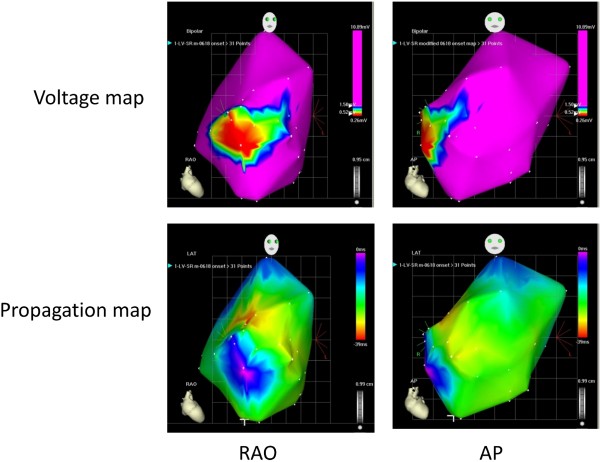
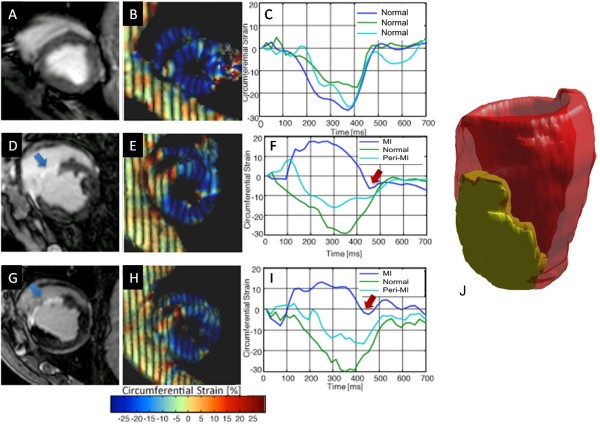
Methods: Cine, gadolinium-contrast, and tagged cardiovascular magnetic resonance (CMR) were performed pre-MI, 9 ± 2 days (early post-MI), and 33 ± 10 days (late post-MI) post-MI in 6 pigs to characterize cardiac morphology, location and extent of MI, and regional mechanics. LV mechanics were assessed by circumferential strain (eC). Electro-anatomic mapping (EAM) was performed within 24 hrs of CMR and prior to sacrifice.
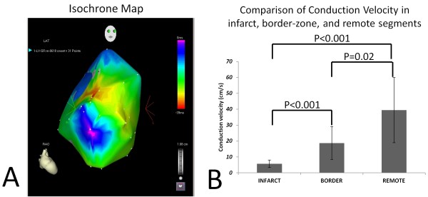
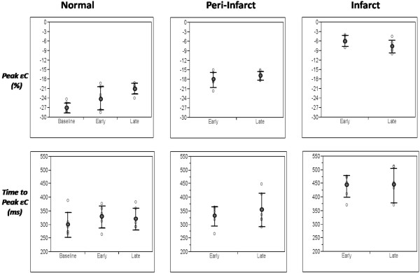
Results: Mean infarct size was 21 ± 4% of LV volume with evidence of post-MI remodeling. Global eC significantly decreased post MI (-27 ± 1.6% vs. -18 ± 2.5% (early) and -17 ± 2.7% (late), p < 0.0001) with no significant change in peri-MI and MI segments between early and late time-points. Time to peak strain (TTP) was significantly longer in MI, compared to normal and peri-MI segments, both early (440 ± 40 ms vs. 329 ± 40 ms and 332 ± 36 ms, respectively; p = 0.0002) and late post-MI (442 ± 63 ms vs. 321 ± 40 ms and 355 ± 61 ms, respectively; p = 0.012). The standard deviation of TTP in 16 segments (SD16) significantly increased post-MI: 28 ± 7 ms to 50 ± 10 ms (early, p = 0.012) to 54 ± 19 ms (late, p = 0.004), with no change between early and late post-MI time-points (p = 0.56). TTP was not related to reduction of segmental contractility. EAM revealed late electrical activation and greatly diminished conduction velocity in the infarct (5.7 ± 2.4 cm/s), when compared to peri-infarct (18.7 ± 10.3 cm/s) and remote myocardium (39 ± 20.5 cm/s).
Conclusions: Mechanical dyssynchrony occurs early after MI and is the result of delayed electrical and mechanical activation in the infarct.
© 2012 Abd-Elmoniem et al; licensee BioMed Central Ltd.
Figures
References
-
- Young JB, Abraham WT, Smith AL, Leon AR, Lieberman R, Wilkoff B, Canby RC, Schroeder JS, Liem LB, Hall S, Wheelan K. Combined cardiac resynchronization and implantable cardioversion defibrillation in advanced chronic heart failure: the MIRACLE ICD Trial. JAMA. 2003;289:2685–2694. doi: 10.1001/jama.289.20.2685. - DOI - PubMed
-
- Cazeau S, Leclercq C, Lavergne T, Walker S, Varma C, Linde C, Garrigue S, Kappenberger L, Haywood GA, Santini M. et al. Effects of multisite biventricular pacing in patients with heart failure and intraventricular conduction delay. N Engl J Med. 2001;344:873–880. doi: 10.1056/NEJM200103223441202. - DOI - PubMed
-
- Aranda JM Jr, Woo GW, Schofield RS, Handberg EM, Hill JA, Curtis AB, Sears SF, Goff JS, Pauly DF, Conti JB. Management of heart failure after cardiac resynchronization therapy: integrating advanced heart failure treatment with optimal device function. J Am Coll Cardiol. 2005;46:2193–2198. doi: 10.1016/j.jacc.2005.03.078. - DOI - PubMed
Publication types
MeSH terms
Substances
LinkOut - more resources
Full Text Sources
Medical
Molecular Biology Databases
