. 2012 Mar;45(3):1129-35.
doi: 10.1016/j.nbd.2011.12.034. Epub 2011 Dec 27.

Loss of circadian clock accelerates aging in neurodegeneration-prone mutants

Affiliations

Loss of circadian clock accelerates aging in neurodegeneration-prone mutants

Natraj Krishnan et al. Neurobiol Dis. 2012 Mar.

Abstract

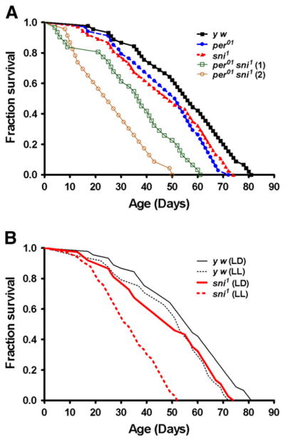
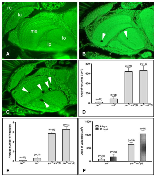
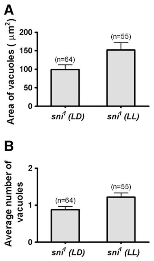
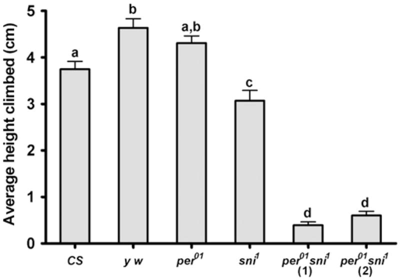
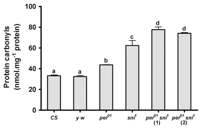
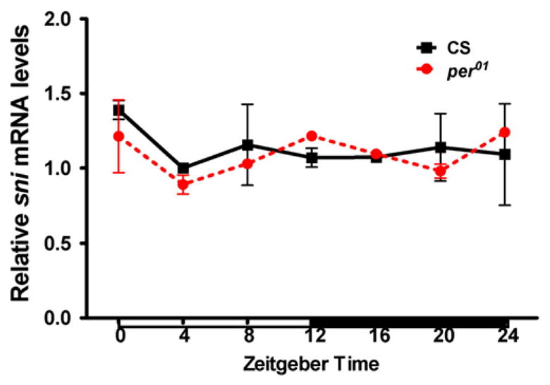
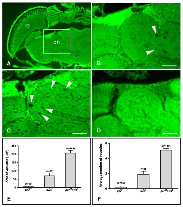
Circadian clocks generate rhythms in molecular, cellular, physiological, and behavioral processes. Recent studies suggest that disruption of the clock mechanism accelerates organismal senescence and age-related pathologies in mammals. Impaired circadian rhythms are observed in many neurological diseases; however, it is not clear whether loss of rhythms is the cause or result of neurodegeneration, or both. To address this important question, we examined the effects of circadian disruption in Drosophila melanogaster mutants that display clock-unrelated neurodegenerative phenotypes. We combined a null mutation in the clock gene period (per(01)) that abolishes circadian rhythms, with a hypomorphic mutation in the carbonyl reductase gene sniffer (sni(1)), which displays oxidative stress induced neurodegeneration. We report that disruption of circadian rhythms in sni(1) mutants significantly reduces their lifespan compared to single mutants. Shortened lifespan in double mutants was coupled with accelerated neuronal degeneration evidenced by vacuolization in the adult brain. In addition, per(01)sni(1) flies showed drastically impaired vertical mobility and increased accumulation of carbonylated proteins compared to age-matched single mutant flies. Loss of per function does not affect sni mRNA expression, suggesting that these genes act via independent pathways producing additive effects. Finally, we show that per(01) mutation accelerates the onset of brain pathologies when combined with neurodegeneration-prone mutation in another gene, swiss cheese (sws(1)), which does not operate through the oxidative stress pathway. Taken together, our data suggest that the period gene may be causally involved in neuroprotective pathways in aging Drosophila.


Figures

Fig. 1
Fig. 1
Loss of circadian rhythms dramatically shortens the lifespan of sni1 mutants. A) Survival curves for y w, per01, sni1, and per01 sni1 double mutant lines. B) Lifespan of sni1 and y w in 12 h light: dark (LD) cycles and constant light (LL), which disrupts the circadian clock function.
Fig. 2
Fig. 2
Interfering with the clock increases neurodegeneration in sni1 mutants. A–C) Paraffin head sections from 9 day-old males (scale bar=25 μm, re=retina, la=lamina, me=medulla, lo=lobula, lp=lobula plate). A) No vacuoles are detectable in the brain of a per01 fly. B) A sni1 fly brain shows a few vacuoles (arrows). C) Brains of per01 sni1 double mutant show increase in the size and number of vacuoles. D) Bar graph showing the mean±SEM area of all vacuoles/brain hemisphere. There is a significant difference between the sni1 and per01 sni1 line 1 (p=2.9×10⁻⁶), and sni1 and per01 sni1 line 2 (p=1.6×10⁻⁵). E) The mean number of vacuoles/brain hemisphere is increased in per01 sni1 compared to sni1 alone [sni1 to per01 sni1 (1): p=8.42×10⁻¹²; sni1 to per01 sni1 (2): p=6.5×10⁻⁸]. F) Comparison of the vacuolization between 9 and 19 day-old flies shows that the phenotype is progressive with age for both sni1 (p=0.036) and per01 sni1 line 1 (p=0.03). D–F) The number of brain hemispheres (n) examined to calculate the average values are indicated on the top of each bar.
Fig. 3
Fig. 3
Disrupting the circadian clock by constant light increases vacuolization in sni1 mutant. A) 9 day-old sni1 flies maintained in constant light (LL) show significant increase in the mean area of vacuoles/brain hemisphere compared to 9 day-old sni1 flies in 12 h light:dark (LD, 12:12) cycles (p=0.024). B) The mean number of vacuoles is also significantly higher in sni1 mutant kept in LL (p=0.018). A–B) The number of brain hemispheres (n) examined to calculate the average values is indicated on the top of each bar.
Fig. 4
Fig. 4
per01 sni1 double mutants show accelerated mobility impairment. Vertical mobility was measured by the RING assay in 10 day-old males of the indicated genotypes. Bars represent mean height climbed (±SEM), based on testing 2 vials per genotype, each containing 25 flies. Bars with different superscripts are significantly different at p<0.01.
Fig. 5
Fig. 5
Oxidative damage in the form of protein carbonyls accumulates to higher levels in per01 sni1 flies. Protein carbonyl levels were measured in heads of 10 day-old males of the indicated genotypes. Both per01 and sni1 single mutants had higher protein carbonyls than their respective CS and y w controls. Protein carbonyls were further elevated in per01 sni1 double mutants, compared to per01 or sni1 single mutants. Bars represent mean carbonyl levels (±SEM), based on testing 3 independent sets of flies each containing 75 flies in 3 technical repeats of 25 flies each. Bars with different superscripts are significantly different at p<0.01.
Fig. 6
Fig. 6
Relative sni mRNA levels are not significantly different between CS and per01 mutants. Expression profile of sni was analyzed by qRT-PCR in heads of flies collected at 4 h intervals in LD, 12:12 cycles. White and black horizontal bars indicate periods of light and darkness, respectively.
Fig. 7
Fig. 7
Loss of per function increases neurodegeneration in sws mutants. A) Paraffin head sections from a 14 day-old sws1 fly show widespread degeneration (arrows) characteristic for this mutant (scale bar=50 μm, re=retina, ol=optic lobes, dn=deutocerebral neuropil). B) Magnification from A (box), showing the deutocerebral neuropil that was used for measurements. C) Age-matched per01 sws1 double mutants show increase in the size and number of vacuoles compared to sws1 single mutants. D) Age-matched y w control does not show vacuoles in this area. B–D) Scale bar=25 μm. E) Bar graph showing significant difference in the mean area of all vacuoles in the deutocerebral neuropil between sws1 and per01 sws1 (p=1.9×10⁻⁸). F) The mean number of vacuoles/brain hemisphere is also significantly higher in the double mutant compared to sws1 alone (p=0.0033). E–F) The number of brain hemispheres (n) examined to calculate average values is indicated on the top of each bar.

References

    1. Allada R, Chung BY. Circadian organization of behavior and physiology in Drosophila. Annu Rev Physiol. 2010;72:605–624. - PMC - PubMed
    1. Antoch MP, et al. Disruption of the circadian clock due to the Clock mutation has discrete effects on aging and carcinogenesis. Cell Cycle. 2008;7:1197–1204. - PMC - PubMed
    1. Beaver LM, et al. Circadian clock regulates response to pesticides in Drosophila via conserved Pdp1 pathway. Toxicol Sci. 2010;115:513–520. - PMC - PubMed
    1. Bettencourt da Cruz A, et al. Disruption of the MAP1B-related protein FUTSCH leads to changes in the neuronal cytoskeleton, axonal transport defects, and progressive neurodegeneration in Drosophila. Mol Biol Cell. 2005;16:2433–2442. - PMC - PubMed
    1. Bettencourt Da Cruz A, et al. Swiss Cheese, a protein involved in progressive neurodegeneration, acts as a noncanonical regulatory subunit for PKA-C3. J Neurosci. 2008;28:10885–10892. - PMC - PubMed

Publication types

MeSH terms