. 2012 May;235(1):162-73.
doi: 10.1016/j.expneurol.2011.12.037. Epub 2011 Dec 29.

Conditioning lesions before or after spinal cord injury recruit broad genetic mechanisms that sustain axonal regeneration: superiority to camp-mediated effects

Affiliations

Conditioning lesions before or after spinal cord injury recruit broad genetic mechanisms that sustain axonal regeneration: superiority to camp-mediated effects

Armin Blesch et al. Exp Neurol. 2012 May.

Abstract

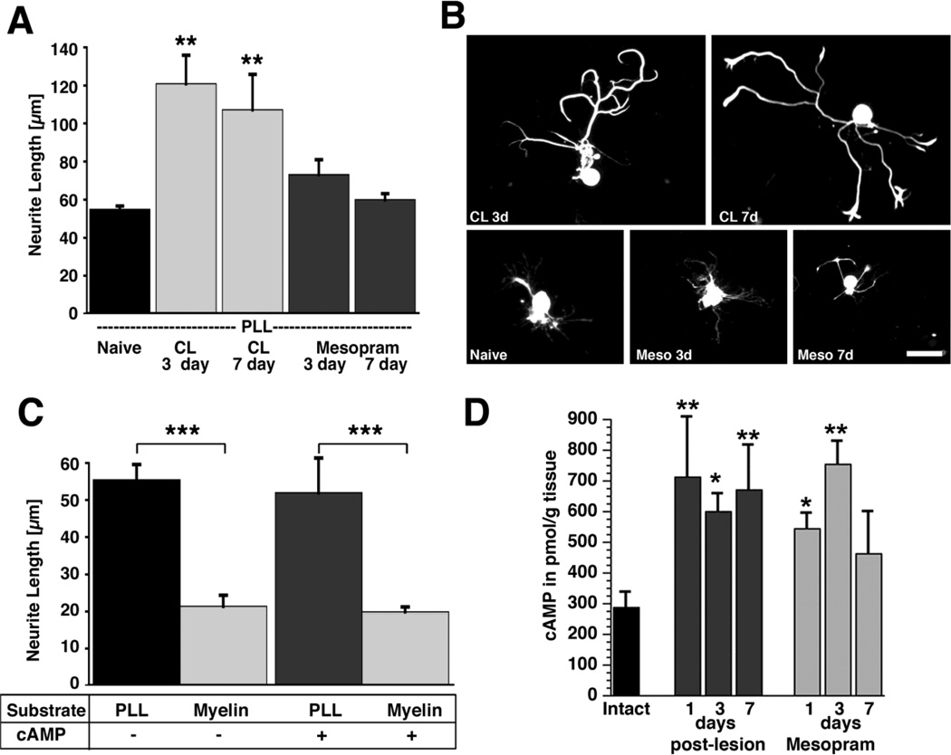
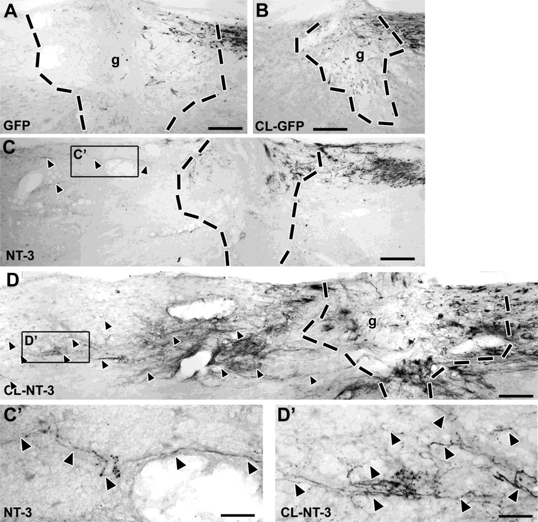
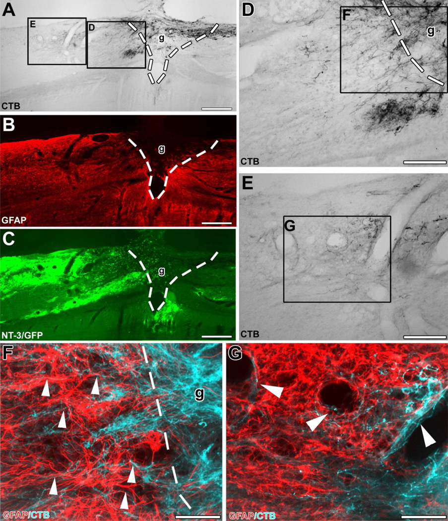
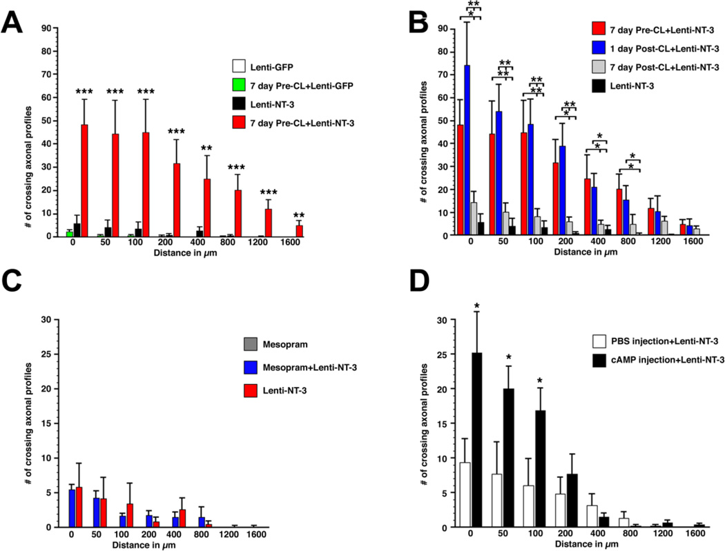
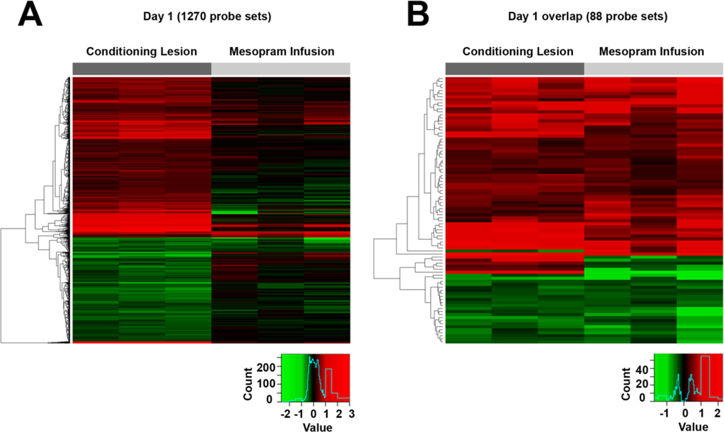
Previous studies indicate that peripheral nerve conditioning lesions significantly enhance central axonal regeneration via modulation of cAMP-mediated mechanisms. To gain insight into the nature and temporal dependence of neural mechanisms underlying conditioning lesion effects on central axonal regeneration, we compared the efficacy of peripheral sciatic nerve crush lesions to cAMP elevations (in lumbar dorsal root ganglia) on central sensory axonal regeneration when administered either before or after cervical spinal cord lesions. We found significantly greater effects of conditioning lesions compared to cAMP elevations on central axonal regeneration when combined with cellular grafts at the lesion site and viral neurotrophin delivery; further, these effects persisted whether conditioning lesions were applied prior to or shortly after spinal cord injury. Indeed, conditioning lesions recruited extensively greater sets of genetic mechanisms of possible relevance to axonal regeneration compared to cAMP administration, and sustained these changes for significantly greater time periods through the post-lesion period. We conclude that cAMP-mediated mechanisms account for only a portion of the potency of conditioning lesions on central axonal regeneration, and that recruitment of broader genetic mechanisms can extend the effect and duration of cellular events that support axonal growth.


Figures

Figure 1
Figure 1. Quantification of in vitro neurite growth and cAMP levels in adult DRG neurons after conditioning lesions and infusion of phosphodiesterase inhibitors
(A) Adult lumbar DRGs (L4–6) were dissected from naïve animals, animals that underwent a sciatic nerve crush 3 or 7 prior to isolation, or animals that received subcutaneous infusions of mesopram for 3 or 7 days prior to isolation. Quantification of neurite length indicates a significant increase in neurite outgrowth on poly-L-lysine (PLL) 3 and 7 days after conditioning lesions (**p<0.01 compared to naive), whereas infusions of the PDE IV inhibitor mesopram have no effect. (B) Examples of NF-200 labeled neurons indicate enhanced growth 3 days (CL 3d) and 7 days (CL 7d) after conditioning lesions but not after mesopram infusion (Meso 3d; Meso 7d). (C) db-cAMP (2 mM) does not increase neurite outgrowth of adult neurons on PLL or myelin. Myelin is strongly inhibitory in the presence and absence of db-cAMP (*** p<0.001 comparing PLL to myelin). Cells were cultivated for 72h and labeled for NF-200 to identify large and medium sized neurons. Data are presented as the means ± SEM of the average neurite length obtained in at least 3 independent experiments. (D) Quantification of cAMP levels in DRGs by ELISA. L4–6 DRGs were dissected from naïve animals, animals that underwent sciatic nerve crush lesions, and animals that received subcutaneous infusions of mesopram for the time indicated. Sciatic nerve lesions result in significant increases in cAMP levels at 1, 3 and 7 days post-lesion. Infusions of mesopram only lead to transient increases in cAMP levels (ANOVA followed by Fischer’s posthoc testing **p<0.01, * p<0.05, compared to naïve controls). Scale bar= 79 µm in (B).
Figure 2
Figure 2. A combination of pre-conditioning lesions and lentiviral NT-3 gene transfer results in significant axonal bridging across the lesion site
(A) CTB-labeled ascending sensory axons fail to bridge across a lesion site filled with BMSC in control animals that received injections of Lenti-GFP. (B) Conditioning lesions combined with control Lenti-GFP injections do not increase the number of bridging axons. (C) Animals that received Lenti-NT-3 rostral to a lesion (without conditioning lesions) show some bridging axons extending only for short distances (quantified in Fig. 4). (D) In contrast, a combination of Lenti-NT-3 and pre-conditioning lesions results in numerous axon bridging beyond the lesion and extension for longer distances. (C’, D‘) Higher magnification of boxed areas in (C) and (D), respectively, showing regenerating CTB-labeled axons (arrowheads) rostral to the lesion site. Rostral is to the left, dorsal to the top. Scale bar= 170 µm in (A–D), 42 µm in (C’, D’). Dashed lines indicate lesion/graft site determined by the absence of GFAP immunolabeling (see Fig. 3).
Figure 3
Figure 3. Ascending sensory axons extend beyond the graft/lesion site towards Lenti-NT-3-transduced cells in an animal that received pre-conditioning lesions and Lenti-NT-3 gene transfer
Triple immunolabeling for (A) CTB to label ascending sensory axons, (B) GFAP to indicate the extent of the lesion/graft, and (C) GFP to label Lenti-NT-3-transduced cells in sagittal spinal cord sections. (D, E) Higher magnification of insets in (A) shows (D) axons growing across the rostral graft (g)/lesion site border (indicated by dashed lines). (E) Numerous axons are present further rostral to the lesion site, shown at higher magnification in (G). (F, G) Double immunolabeling for CTB-labeled axons (pseudocolored blue) and GFAP (red) at the (F) rostral host/graft interface (indicated by dashed lines) and (G) in the host spinal cord beyond the lesion. Bridging axons were often found to (F) orient along GFAP-labeled processes (arrowheads) and (G) were occasionally associated with blood vessels beyond the lesion site. Rostral is to the left, dorsal to the top. Scale bars 424 µm in (A–C), 170 µm in (D, E), 85 µm in (F, G).
Figure 4
Figure 4. Quantification of axons bridging across a C3 lesion site filled with bone marrow stromal cells in animals that received pre- or post-conditioning lesions, infusions of mesopram or db-cAMP injections
(A) Pre-conditioning lesions in combination with Lenti-NT-3 gene transfer (7 day Pre-CL+Lenti-NT-3) significantly increased the number of axons crossing a C3 dorsal funiculus lesion compared to animals that received only Lenti-NT-3, only Lenti-GFP, or a combination of Lenti-GFP/conditioning lesions (7 day Pre-CL-Lenti-GFP). The number of axons crossing the rostral host graft interface (0 µm) or a virtual line 50, 100, 200, 400, 800, 1200 and 1600 µm beyond the lesion site was quantified in a series of 1 out of 7 sections. (ANOVA followed by Fischer’s posthoc analysis *** p<0.001, ** p<0.01 comparing 7 day Pre-CL+Lenti-NT-3 to all other groups). (B) Axonal bridging after 1- or 7-day post-conditioning lesions in combination with Lenti-NT-3 gene transfer. The number of bridging axons in animals that received post-conditioning lesions 1 day after central lesions is not significantly different at any distance examined from animals that received pre-conditioning lesions 7 day before central lesions. Priming neurons by peripheral lesions 7 days after spinal cord lesions resulted in significantly fewer axons bridging beyond the lesion site. Data from pre-conditioned animals in combination with Lenti-NT-3 from (A) are included for clarity. (ANOVA followed by Fischer’s posthoc testing **p<0.01; * p<0.05). (C, D) Increases in cAMP levels result in only short-distance axon growth beyond the lesion. (C) Animals received infusions of the phosphodiesterase inhibitor mesopram starting one week prior to C3 dorsal column lesions, BMSC grafts, and Lenti-NT-3 or Lenti-GFP injections. Infusions of mesopram without NT-3 delivery failed to result in any axonal bridging; the combination of mesopram infusion with Lenti-NT-3 delivery did not further increase the number of bridging axons compared to Lenti-NT-3 delivery alone. Animals with Lenti-NT-3 injections alone, quantified in (A), are included for comparison. (D) Injections of dibutyryl-cAMP into L4 and L5 DRGs one day after spinal cord lesions and Lenti-NT-3 delivery increase axonal bridging only up to 100 µm beyond the rostral host/graft interface, when compared to animals injected with PBS and Lenti-NT-3 as controls (unpaired t-test *p<0.05). Data represent raw counts in one out of seven sections.
Figure 5
Figure 5. Heat maps depicting fold changes in gene expression one day following conditioning lesions and infusions of the phosphodiesterase inhibitor mesopram
(A) A total of 898 probe sets are differentially expressed in DRGs after conditioning lesions (pFDR<0.05; change of > 20%) with limited overlap to the genes differentially expressed after mesopram infusion. (B) Gene expression is altered in the same direction for nearly all significantly changed probe sets overlapping between both groups. All comparisons are to DRGs of naïve control animals (see also Supplementary Data). Each column represents data from one array.

References

    1. Alto LT, Havton LA, Conner JM, Hollis Ii ER, Blesch A, Tuszynski MH. Chemotropic guidance facilitates axonal regeneration and synapse formation after spinal cord injury. Nat Neurosci. 2009;12:1106–1113. - PMC - PubMed
    1. Andersen PL, Webber CA, Kimura KA, Schreyer DJ. Cyclic AMP prevents an increase in GAP-43 but promotes neurite growth in cultured adult rat dorsal root ganglion neurons. Exp Neurol. 2000;166:153–165. - PubMed
    1. Azizi SA, Stokes D, Augelli BJ, DiGirolamo C, Prockop DJ. Engraftment and migration of human bone marrow stromal cells implanted in the brains of albino rats--similarities to astrocyte grafts. Proc Natl Acad Sci U S A. 1998;95:3908–3913. - PMC - PubMed
    1. Benjamini Y, Hochberg Y. Controlling the false discovery rate: a practical and powerful approach to multiple testing. Journal of the Royal Statistical Society. 1995;57:289–300.
    1. Bethea JR, Dietrich WD. Targeting the host inflammatory response in traumatic spinal cord injury. Curr Opin Neurol. 2002;15:355–360. - PubMed

Publication types