. 2012 Jun;46(6):748-56.
doi: 10.1165/rcmb.2011-0224OC. Epub 2012 Jan 6.

Role of endoplasmic reticulum stress in age-related susceptibility to lung fibrosis

Affiliations

Role of endoplasmic reticulum stress in age-related susceptibility to lung fibrosis

Edilson Torres-González et al. Am J Respir Cell Mol Biol. 2012 Jun.

Abstract

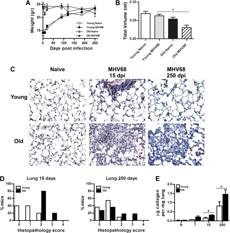
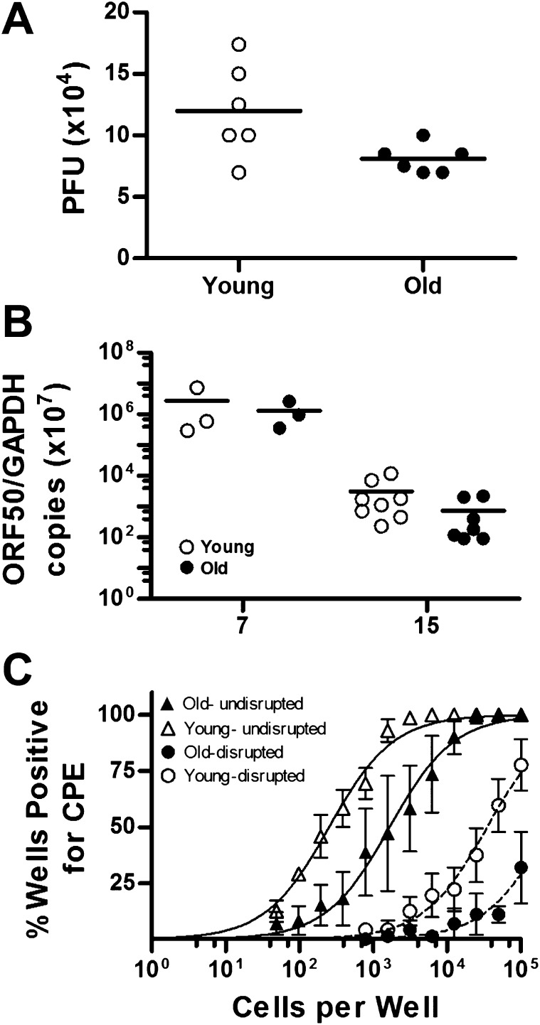
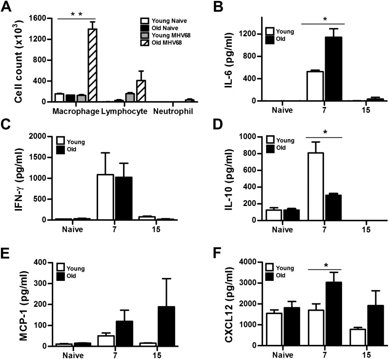
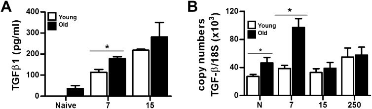
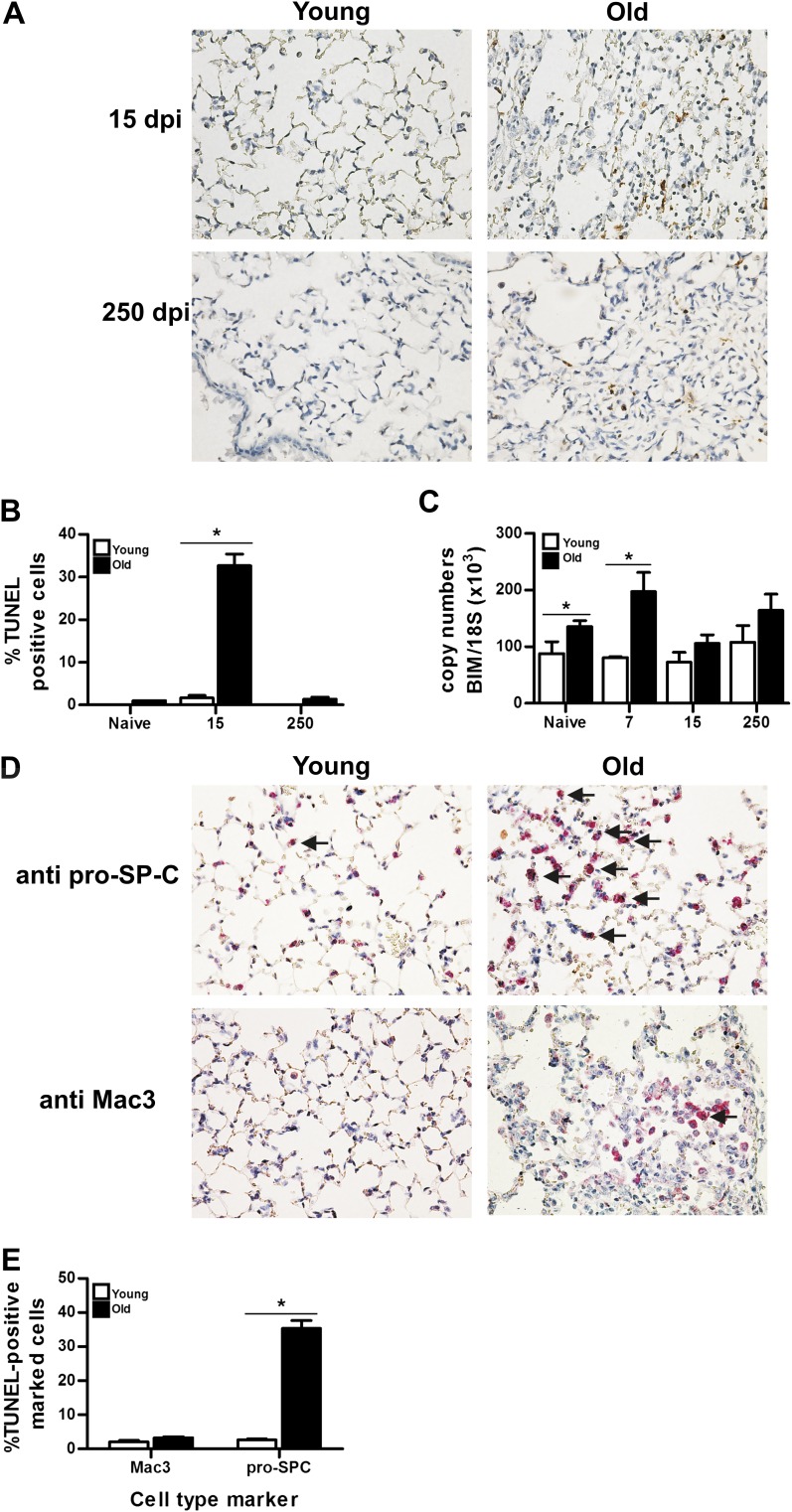
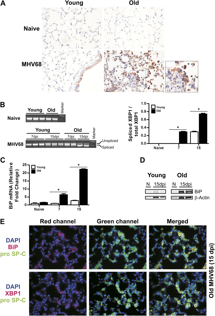
The incidence of idiopathic pulmonary fibrosis (IPF) increases with age. The mechanisms that underlie the age-dependent risk for IPF are unknown. Based on studies that suggest an association of IPF and γherpesvirus infection, we infected young (2-3 mo) and old (≥18 mo) C57BL/6 mice with the murine γherpesvirus 68. Acute murine γherpesvirus 68 infection in aging mice resulted in severe pneumonitis and fibrosis compared with young animals. Progressive clinical deterioration and lung fibrosis in the late chronic phase of infection was observed exclusively in old mice with diminution of tidal volume. Infected aging mice showed higher expression of transforming growth factor-β during the acute phase of infection. In addition, aging, infected mice showed elevation of proinflammatory cytokines and the fibrocyte recruitment chemokine, CXCL12, in bronchoalveolar lavage. Analyses of lytic virus infection and virus reactivation indicate that old mice were able to control chronic infection and elicit antivirus immune responses. However, old, infected mice showed a significant increase in apoptotic responses determined by in situ terminal deoxynucleotidyl transferase dUTP nick end labeling assay, levels of caspase-3, and expression of the proapoptotitc molecule, Bcl-2 interacting mediator. Apoptosis of type II lung epithelial cells in aging lungs was accompanied by up-regulation of endoplasmic reticulum stress marker, binding immunoglobulin protein, and splicing of X-box-binding protein 1. These results indicate that the aging lung is more susceptible to injury and fibrosis associated with endoplasmic reticulum stress, apoptosis of type II lung epithelial cells, and activation of profibrotic pathways.


Figures

Figure 1.
Figure 1.
Severe clinical disease and fibrosis in murine γherpesvirus (MHV) 68–infected aging mice. (A) Weight loss data are presented as total body weight. More severe illness was observed in aging infected mice (number of mice: 5–13 per group and time point). Data are representative of three different experiments. (B) Lung function determined by the measurement of tidal volume using a whole-body plethysmograph. Measurements were performed at Day 120 after infection (number of animals: 3–5 per group; *P < 0.05). (C) Masson trichrome staining of lung sections from 3-month-old (young) and >18-mo-old (old) C57BL/6 mice infected with MHV68 at indicated times after infection. Collagen deposition is shown in blue. Notice loss of normal lung architecture of the lung in old infected mice associated with higher interstitial collagen deposition. Young mice only show collagen deposition around blood vessels at Day 250 after infection. (D) Semiquantitative morphometric analysis of lung histopathology in virus-infected young and old mice at the indicated time after infection. Old infected mice showed higher pathology scores corresponding to pneumonitis and thickening of the interalveolar septa compared with young animals (n = 5–8). (E) Normalized lung collagen content based on hydroxyproline microplate assay at 7, 15, and 250 days after infection (dpi) (*P < 0.05; n = 3).
Figure 2.
Figure 2.
Control of chronic virus infection in young and aging mice. (A) Acute replication in the lungs of young and old C57BL/6 mice at Day 7 after infection. Lungs were harvested, disrupted, and titered on NIH 3T12 cells by plaque assay. Data are shown as log10 titer, and the bar indicates the geometric mean titer. (B) Quantitative PCR analysis of virus load of individual lungs from MHV68-infected young and old C57BL/6 mice at 7 and 15 dpi. Viral load was determined as the levels of viral DNA (targeting the using open reading frame [ORF] 50 region) normalized by cellular DNA (targeting glyceraldehyde 3-phosphate dehydrogenase). Bars represent geometric mean. (C) Reactivation of MHV68 in spleens from infected young and old mice was assessed using standard limiting dilution analyses at 15 dpi (n = 3–4 mice). Serial dilutions of bulk intact or disrupted splenocytes were plated on monolayers of mouse embryonic fibroblasts (MEFs). The presence of reactivating virus was determined by the presence of cytopathic effect (CPE). CPE observed in the disrupted splenocytes indicate preformed infectious virus. Symbols represent the mean percentage of wells positive for CPE (±SEM). Curve fit lines were derived from nonlinear regression analysis. PFU, plaque-forming units.
Figure 3.
Figure 3.
Higher levels of proinflammatory cytokines and chemokines in MHV68-infected aging mice. (A) Differential cell counts in the bronchoalveolar lavage (BAL) of naive and infected young and old mice at 15 dpi. (**P < 0.01; n = 8). (BF) IL-6, IFN-γ, IL-10, and MCP-1 levels were measured in BAL fluid from naive and MHV68-infected young and old mice at the indicated time points after infection, whereas CXCL12 was measured in lung tissue. Bars represent means (±SEM) (*P < 0.05; n = 5 per group).
Figure 4.
Figure 4.
High expression of transforming growth factor (TGF)-β in naive and virus-infected aging mice. (A) Active TGF-β levels were measured in BAL fluid from naive and MHV68 infected young and old mice at 7 and 15 dpi (n = 3). (B) Quantitative RT-PCR analysis for TGF-β transcripts in the lungs of naive and infected young and old mice at different time point after infection (*P < 0.05; n = 4).
Figure 5.
Figure 5.
Apoptosis in type II epithelial cells from infected old C57BL/6 mice. (A) Representative images from in situ terminal deoxynucleotidyl transferase dUTP nick end labeling (TUNEL) assay in lung sections from young and old mice at 15 and 250 dpi. Magnification, 40×. (B) Semiquantitative analyses in high-power field show a greater percentage of TUNEL positive cells in old infected mice (n = 3). (C) Quantitative RT-PCR analysis for Bim transcripts in the lungs of naive and infected young and old mice at different time point after infection (n = 4). (D) Macrophage and type II lung epithelial cell death was evaluated in paraffin-embedded tissue sections from 15 dpi using TUNEL assay (brown). Macrophages and type II lung epithelial cells were identified using Mac3 and pro–surfactant-C antibodies (red). Higher number of apoptotic type II lung epithelial cells (arrows) was found in old infected mice. (E) Semi-quantitative analyses in high-power field show a greater percentage of double stained cells (TUNEL positive nucleus + cell type marker) in old infected mice (n = 4). SP-C, surfactant protein-C.
Figure 6.
Figure 6.
Endoplasmic reticulum (ER) stress in lung epithelial cells from infected old C57BL/6 mice. (A) Immunohistochemistry analyses of X-box–binding protein (XBP) 1 expression in lung sections of young and old mice at Day 15 of mock or MHV68 infection. Notice abundant positive staining in old MHV68-infected mice. (B) Determination of mRNA levels of unspliced and spliced XBP1 in lung samples of naive and MHV68-infected mice at Days 7 and 15 after infection. Ratio of spliced versus total XBP1 mRNA of samples is shown on the right. (C) Quantitative RT-PCR analysis for binding immunoglobulin protein (BiP) transcripts in the lungs of naive and infected young and old mice at different time point after infection (n = 3–4; *P < 0.001). (D) Immunoblot assay of lung lysates from naive (N) and infected young and old C57BL/6 mice at the indicated time point after infection using anti-BiP antibody. Blot was stripped and reprobed with an anti–β-actin antibody as a loading control. (E) Dual immunofluorescent staining in lung sections of aging mice infected at 15 dpi for pro–SP-C (green) and the markers of ER stress BiP (red) and XBP1 (red). Yellow cells in the merge column indicate type II cells supporting ER stress. Nuclei were visualized by 4′,6-diamidino-2-phenylindole staining (blue). SP-C, surfactant protein-C.

References

    1. Raghu G, Nyberg F, Morgan G. The epidemiology of interstitial lung disease and its association with lung cancer. Br J Cancer 2004;91 Suppl 2:S3–S10 - PMC - PubMed
    1. Castriotta RJ, Eldadah BA, Foster WM, Halter JB, Hazzard WR, Kiley JP, King TE, Jr, Horne FM, Nayfield SG, Reynolds HY, et al. Workshop on idiopathic pulmonary fibrosis in older adults. Chest 2010;138:693–703 - PMC - PubMed
    1. Stewart JP, Egan JJ, Ross AJ, Kelly BG, Lok SS, Hasleton PS, Woodcock AA. The detection of Epstein-Barr virus DNA in lung tissue from patients with idiopathic pulmonary fibrosis. Am J Respir Crit Care Med 1999;159(4 Pt 1):1336–1341 - PubMed
    1. Egan JJ, Woodcock AA, Stewart JP. Viruses and idiopathic pulmonary fibrosis. Eur Respir J 1997;10:1433–1437 - PubMed
    1. Tang YW, Johnson JE, Browning PJ, Cruz-Gervis RA, Davis A, Graham BS, Brigham KL, Oates JA, Jr, Loyd JE, Stecenko AA. Herpesvirus DNA is consistently detected in lungs of patients with idiopathic pulmonary fibrosis. J Clin Microbiol 2003;41:2633–2640 - PMC - PubMed

Publication types

Substances