. 2012 Jun;6(2):232-43.
doi: 10.1007/s12105-011-0328-1. Epub 2012 Jan 7.

Cytoplasmic ezrin and moesin correlate with poor survival in head and neck squamous cell carcinoma

Affiliations

Cytoplasmic ezrin and moesin correlate with poor survival in head and neck squamous cell carcinoma

Nicolas F Schlecht et al. Head Neck Pathol. 2012 Jun.

Abstract

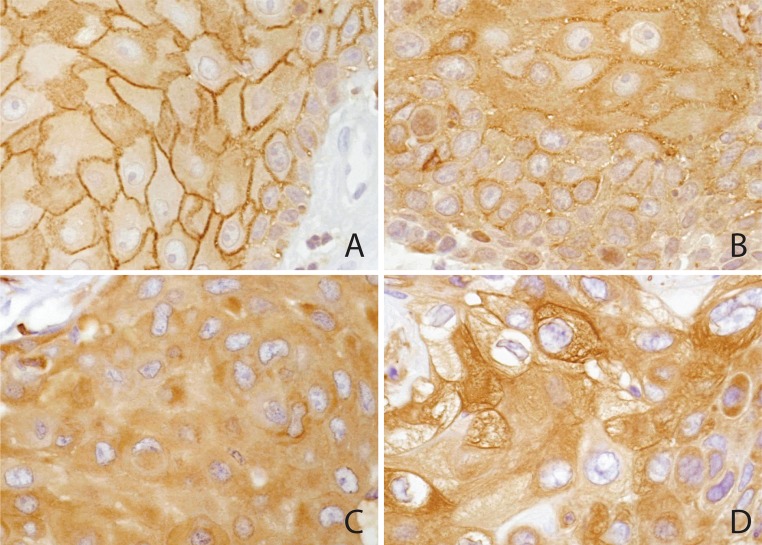
Members of the 4.1 superfamily of proteins, including ezrin, moesin, merlin, and willin regulate many normal physiologic processes such as cellular shape, motility, and proliferation. In addition, they contribute both to tumor development and tumor progression. We reported previously that strong cytoplasmic ezrin expression was independently associated with poorer patient survival. One hundred and thirty-one histologically confirmed primary head and neck squamous cell carcinomas were examined prospectively for cancer progression and survival at a large health care center in the Bronx, NY, USA. Immunohistochemical analysis of ezrin, moesin, merlin, and willin expression in tissue microarray samples of primary head and neck squamous cell carcinoma revealed a significant association of increased cytoplasmic ezrin with poor cancer survival. Global RNA analyses suggest that cancers with high cytoplasmic ezrin have a more invasive phenotype. This study supports our previous findings associating cytoplasmic ezrin with more aggressive behavior and poorer outcome and indicates the need for a multi-institutional study to validate the use of cytoplasmic ezrin as a biomarker for treatment planning in head and neck squamous cell carcinoma.


Figures

Fig. 1
Fig. 1
Cytoplasmic ezrin. a Ezrin expression in normal squamous mucosa. There is strong staining of the cell membranes of the stratum spinosum, but only faint uniform staining of the cytoplasm. Basal cells at the stromal interface are negative. b Squamous cell carcinoma demonstrating weak (+1) cytoplasmic ezrin localization. Prominent membrane localization is accentuated in the interior of the cell group, recapitulating the stratification of normal epithelium. c Squamous cell carcinoma demonstrating moderate (+2) cytoplasmic ezrin expression, with reduced membrane localization. d Squamous cell carcinoma demonstrating strong (+3) cytoplasmic ezrin expression. Many cells have intensely staining cytoplasmic filaments, while membrane localization is minimal. Two pathologists (MBG, DB), blinded to outcome and tissue source, read the microarrays and achieved a consensus regarding staining pattern and intensity. The level of cytoplasmic staining was scored according to the strongest intensity that comprised at least 10% of each core on a scale of 0–3. Mean staining scores were then calculated across multiple core replicates from the same primary tumor biopsy or resection specimen
Fig. 2
Fig. 2
Order of graphs clockwise from top left: a Ezrin expression; b Moesin expression, c Willin expression, and d Merlin expression. Survival time displayed in months truncated at 60 months for illustration. Censored cases shown by tick marks at last recorded follow-up
Fig. 3
Fig. 3
Order of graphs clockwise from top left: a Ezrin expression; b Moesin expression, c Willin expression, and d Merlin expression. Time to first LRR (or distant metastasis) displayed in months truncated at 60 months for illustration. Censored cases shown by tick marks at last recorded follow-up

References

    1. Jemal A, Bray F, Center MM, Ferlay J, Ward E, Forman D. Global cancer statistics. CA Cancer J Clin. 2011;61:69–90. doi: 10.3322/caac.20107. - DOI - PubMed
    1. Belbin TJ, Singh B, Smith RV, et al. Molecular profiling of tumor progression in head and neck cancer. Arch Otolaryngol Head Neck Surg. 2005;131:10–18. doi: 10.1001/archotol.131.1.10. - DOI - PubMed
    1. Fehon RG, McClatchey AI, Bretscher A. Organizing the cell cortex: the role of ERM proteins. Natl Rev Mol Cell Biol. 2010;11:276–287. doi: 10.1038/nrm2866. - DOI - PMC - PubMed
    1. Sudol M, Harvey KF. Modularity in the Hippo signaling pathway. Trends Biochem Sci. 2010;35:627–633. doi: 10.1016/j.tibs.2010.05.010. - DOI - PubMed
    1. Bao Y, Hata Y, Ikeda M, Withanage K. Mammalian Hippo pathway: from development to cancer and beyond [in process citation] J Biochem. 2011;149:361–379. doi: 10.1093/jb/mvr021. - DOI - PubMed

Publication types

MeSH terms