. 2012 Apr 15;21(8):1808-23.
doi: 10.1093/hmg/ddr613. Epub 2012 Jan 6.

Mutations in mouse Ift144 model the craniofacial, limb and rib defects in skeletal ciliopathies

Affiliations

Mutations in mouse Ift144 model the craniofacial, limb and rib defects in skeletal ciliopathies

Alyson Ashe et al. Hum Mol Genet. .

Abstract

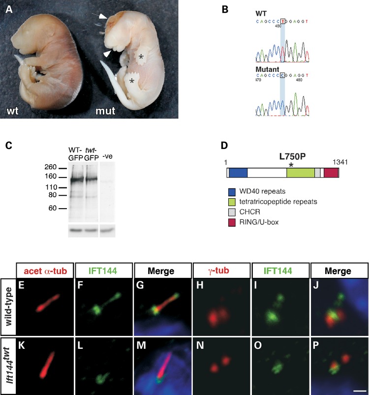
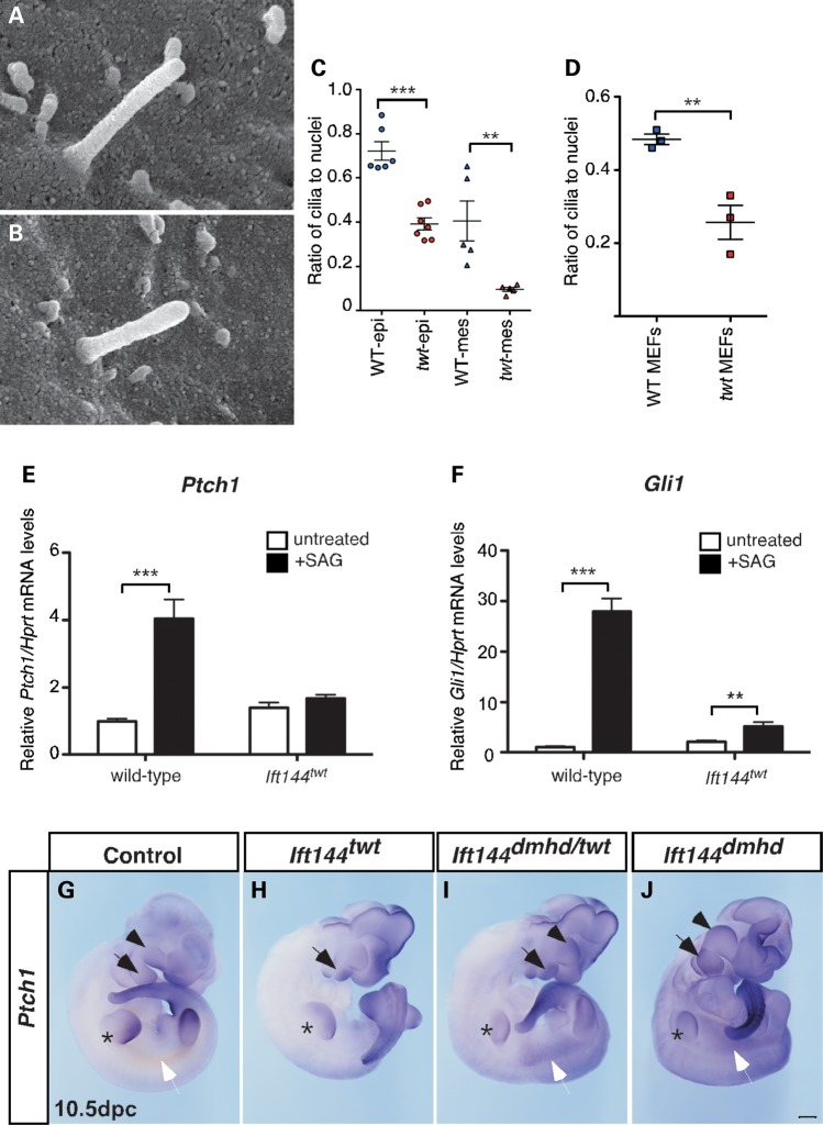
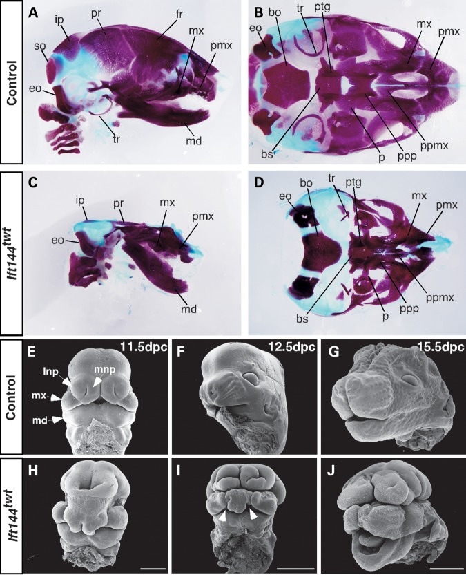
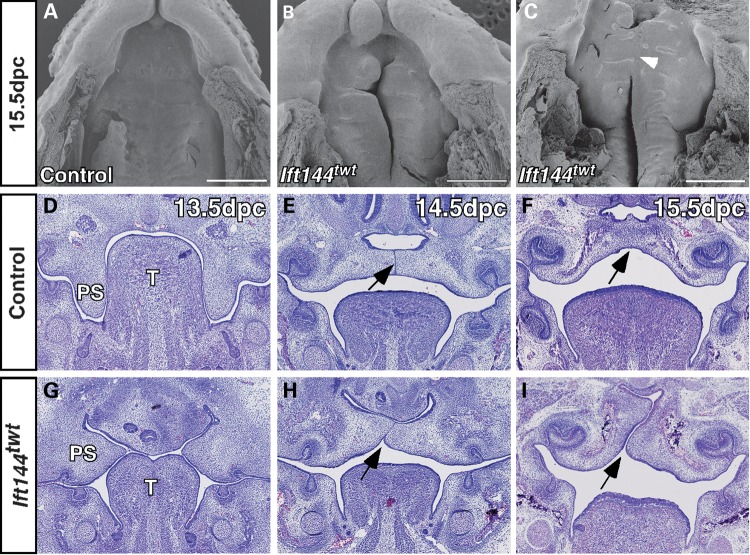
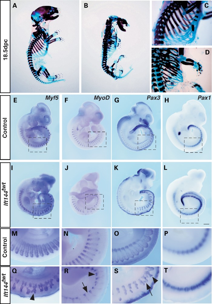
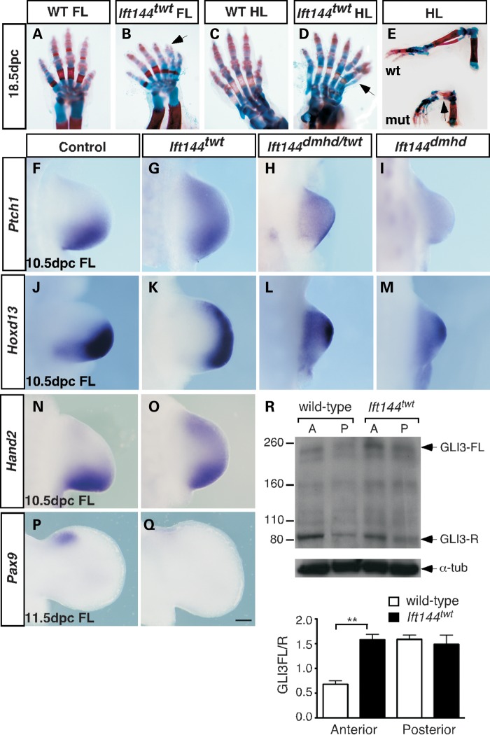
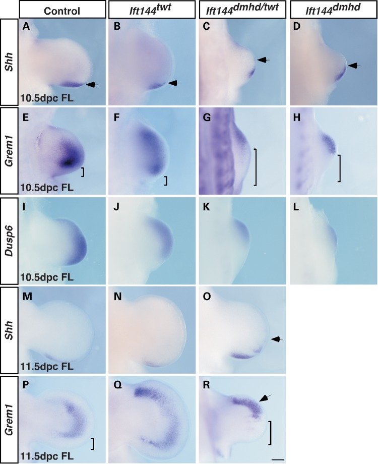
Mutations in components of the intraflagellar transport (IFT) machinery required for assembly and function of the primary cilium cause a subset of human ciliopathies characterized primarily by skeletal dysplasia. Recently, mutations in the IFT-A gene IFT144 have been described in patients with Sensenbrenner and Jeune syndromes, which are associated with short ribs and limbs, polydactyly and craniofacial defects. Here, we describe an N-ethyl-N-nitrosourea-derived mouse mutant with a hypomorphic missense mutation in the Ift144 gene. The mutant twinkle-toes (Ift144(twt)) phenocopies a number of the skeletal and craniofacial anomalies seen in patients with human skeletal ciliopathies. Like other IFT-A mouse mutants, Ift144 mutant embryos display a generalized ligand-independent expansion of hedgehog (Hh) signalling, in spite of defective ciliogenesis and an attenuation of the ability of mutant cells to respond to upstream stimulation of the pathway. This enhanced Hh signalling is consistent with cleft palate and polydactyly phenotypes in the Ift144(twt) mutant, although extensive rib branching, fusion and truncation phenotypes correlate with defects in early somite patterning and may reflect contributions from multiple signalling pathways. Analysis of embryos harbouring a second allele of Ift144 which represents a functional null, revealed a dose-dependent effect on limb outgrowth consistent with the short-limb phenotypes characteristic of these ciliopathies. This allelic series of mouse mutants provides a unique opportunity to uncover the underlying mechanistic basis of this intriguing subset of ciliopathies.


Figures

Figure 1.
Figure 1.
The twt mouse has a causative mutation in the Ift144 gene. (A) Whole wild-type (left) and mutant (right) embryos at 17.5 dpc showing exencephaly (exposed brain, arrow), polydactyly (asterisks) and craniofacial abnormalities, including protruding tongue (arrowhead). (B) A homozygous T-to-C transition at position 2249 of the Ift144 gene causes a leucine-to-proline substitution at amino acid position 750 of the protein (D). (C) A mutant expression construct produces stable exogenous protein at a similar size to the wild-type construct. The negative lane represents protein from untransfected cells; the lower panel shows a tubulin loading control. (EJ) In most wild-type-ciliated MEFs (∼89%), endogenous IFT144 is enriched at the base and tip of the cilium, and appears in a punctate pattern along the axoneme, as shown by co-localization with acetylated α-tubulin at the axoneme (E–G) and staining close to γ-tubulin which marks the basal body (H–J). In Ift144twt MEFs, IFT144 appears preferentially restricted to the base of the cilium (∼78% of ciliated cells analysed; KP), as confirmed by staining close to γ-tubulin (N–P). Scale bar: 1 µm.
Figure 2.
Figure 2.
Ift144twt embryos have fewer ciliated cells and altered Hh signalling. (A and B) SEM revealed no obvious consistent morphological differences in primary cilia projecting from the dorsal surface of the limb of wild-type (A) and Ift144twt mutant (B) embryos. (C and D) Whole-mount IF analysis for ARL13b to mark the axoneme revealed fewer ciliated epithelial and mesenchymal cells in Ift144twt mutant limbs (C) and MEFs (D) compared with wild-type. (E and F) MEFs from wild-type and Ift144twt mutant embryos were exposed to the SMO agonist SAG and the level of Hh signalling assessed by qRT-PCR analysis of Ptch1 (E) and Gli1 (F). The response of Ift144twt MEFs to SAG stimulation was reduced relative to wild-type MEFs. (GJ) WISH reveals enhanced expression of Ptch1 in 10.5 dpc Ift144 mutants. Arrowhead, maxilla; black arrow, mandible; asterisk, forelimb; white arrow, inter-limb flank mesenchyme Scale bar: 500 µm. P-values in (C)–(F) are based on Student's t-test (***P < 0.001;**P < 0.01). Error bars show standard error of the mean.
Figure 3.
Figure 3.
Craniofacial phenotype of Ift144twt embryos. (AD) Skulls of 18.5 dpc control (A and B) and Ift144twt (C and D) embryos stained for bone (Alizarin red) and cartilage (Alcian blue). (EJ) SEM analysis of control (E–G) and Ift144twt mutant (H–J) heads at a range of gestational stages. White arrowheads in (I) mark the bilateral facial cleft in 12.5 dpc Ift144twt embryos. bo, basioccipital; bs, basisphenoid; eo, exoccipital; fr, frontal; ip, interparietal; lnp, lateral nasal process; md, mandible; mx, maxilla; mnp, medial nasal process; p, palatine; pmx, premaxilla; ppmx, palatal process maxilla; ppp, palatal process palatine; pr, parietal; ptg, pterygoid; so, supraoccipital; tr, tympanic ring. Scale bars: (E and H): 500 µm; (F, G, I and J): 1 mm.
Figure 4.
Figure 4.
Ift144twt embryos display cleft palate. (AC) SEM analysis of 15.5 dpc embryonic heads with the jaw removed revealed a cleft palate in Ift144twt embryos. One sample (C) showed some evidence of limited anterior fusion (arrowhead). (DI) Histological analysis of frontal sections through the head of control (D–F), and Ift144twt mutant embryos (G–I) at a range of gestational stages. Mutant palatal shelves do not grow vertically alongside the tongue at 13.5 dpc (D and G), and do not fuse correctly despite often closely abutting one another (E, F, H and I). The arrow marks the MES in (E), its normal removal in (F) and the failure to form correctly in (H) and (I). ps, palatal shelf; t, tongue. Scale bar: 500 µm.
Figure 5.
Figure 5.
Ift144twt embryos display skeletal defects due to altered somite patterning. (AD) 18.5 dpc control (A and C) and Ift144twt mutant (B and D) embryos stained for bone (Alizarin red) and cartilage (Alcian blue). Ift144twt embryos display truncated, fused and branched ribs. (ET) WISH analysis of markers of somite compartments in control (E–H) and Ift144twt mutant (I–L) 10.5 dpc whole embryos. (M–T) Higher magnification of boxed regions in (E–L). Arrowheads show the regions' altered expression relative to controls. Scale bar: 500 µm.
Figure 6.
Figure 6.
The forelimbs of Ift144 mutants display a dose-dependent effect on patterning. (AE) Limbs of wild-type (A, C and E—top) and Ift144twt mutant (B, D and E—bottom) 18.5 dpc embryos stained for bone (Alizarin red) and cartilage (Alcian blue). Ift144twt mutant limbs display preaxial polydactyly (arrow in D) with branching at the digit tips (Arrow in C), and tibial agenesis (arrow in E). (FQ) WISH analysis of a range of markers in the forelimb. At 10.5 dpc, Ptch1 expression is expanded into the anterior of Ift144twt (G), Ift144dmhd/twt (H) and Ift144dmhd (I) mutant forelimbs (FL) compared with control (F). Hoxd13 expression is expanded anteriorly in Ift144twt (K), Ift144dmhd/twt (L) and Ift144dmhd (M) 10.5 dpc mutant forelimbs (FL) compared with control (J). Hand2 expression is upregulated in the anterior of the 10.5 dpc Ift144twt (O) forelimb compared with control (N). The control Pax9 anterior expression domain (P) is undetectable in Ift144twt (Q) mutant limbs at 11.5 dpc (R) IB revealed a subtle increase in the GLI3FL/R ratio specifically in the anterior (A) but not the posterior (P) 11.5 dpc Ift144twt limb. Quantification is based on three independent blots using the same protein samples from pooled limbs. P-value is based on Student's t-test (**P = 0.0022). Error bars show standard error of the mean. Limbs in (A)–(D) are oriented anterior to the right; (F)–(Q), anterior to the top. Scale bar: 200 µm.
Figure 7.
Figure 7.
The SHH–GREM1–FGF signalling loop is prematurely disrupted in Ift144 mutant forelimbs. (AD, MO) Shh expression is unaltered in Ift144twt forelimbs at 10.5 dpc (B) and 11.5 dpc (N) relative to controls (A and M). Shh expression is expanded along the distal periphery in Ift144dmhd/twt (C) and Ift144dmhd (D) forelimbs at 10.5 dpc, and in Ift144dmhd/twt at 11.5 dpc (O). Arrows in (A)–(D) and (O) mark the anterior limit of Shh expression. (EH, PR) Grem1 expression is expanded anteriorly in 10.5 dpc (F) and 11.5 dpc (Q) Ift144twt forelimbs compared with controls (E, P). In both Ift144dmhd/twt (G) and Ift144dmhd (H) 10.5 dpc forelimbs, Grem1 expression is expanded anteriorly but reduced in the posterior relative to control (E). A similar pattern is seen in Ift144dmhd/twt forelimbs at 11.5 dpc (R). Square brackets in (E)–(H), (P), (R) denote the region of the posterior limb devoid of Grem1 expression. (IL) Dusp6 expression suggests attenuated FGF signalling in the posterior of Ift144dmhd/twt (K) and Ift144dmhd (L) and, to a lesser extent, Ift144twt (J) forelimbs at 10.5 dpc relative to control (I). Limbs are oriented anterior to the top. Scale bar: 200 µm.

References

    1. Goetz S.C., Anderson K.V. The primary cilium: a signalling centre during vertebrate development. Nat. Rev. Genet. 2010;11:331–344. - PMC - PubMed
    1. Waters A.M., Beales P.L. Ciliopathies: an expanding disease spectrum. Pediatr. Nephrol. 2011;26:1039–1056. - PMC - PubMed
    1. Novarino G., Akizu N., Gleeson J.G. Modeling human disease in humans: the ciliopathies. Cell. 2011;147:70–79. - PMC - PubMed
    1. Beales P.L., Bland E., Tobin J.L., Bacchelli C., Tuysuz B., Hill J., Rix S., Pearson C.G., Kai M., Hartley J., et al. IFT80, which encodes a conserved intraflagellar transport protein, is mutated in Jeune asphyxiating thoracic dystrophy. Nat. Genet. 2007;39:727–729. - PubMed
    1. Mill P., Lockhart P.J., Fitzpatrick E., Mountford H.S., Hall E.A., Reijns M.A., Keighren M., Bahlo M., Bromhead C.J., Budd P., et al. Human and mouse mutations in WDR35 cause short-rib polydactyly syndromes due to abnormal ciliogenesis. Am. J. Hum. Genet. 2011;88:508–515. - PMC - PubMed

Publication types

MeSH terms