. 2012 Jun;90(6):707-18.
doi: 10.1007/s00109-011-0851-2. Epub 2012 Jan 7.

Gastrin inhibits a novel, pathological colon cancer signaling pathway involving EGR1, AE2, and P-ERK

Affiliations

Gastrin inhibits a novel, pathological colon cancer signaling pathway involving EGR1, AE2, and P-ERK

Ling-Jun Song et al. J Mol Med (Berl). 2012 Jun.

Abstract

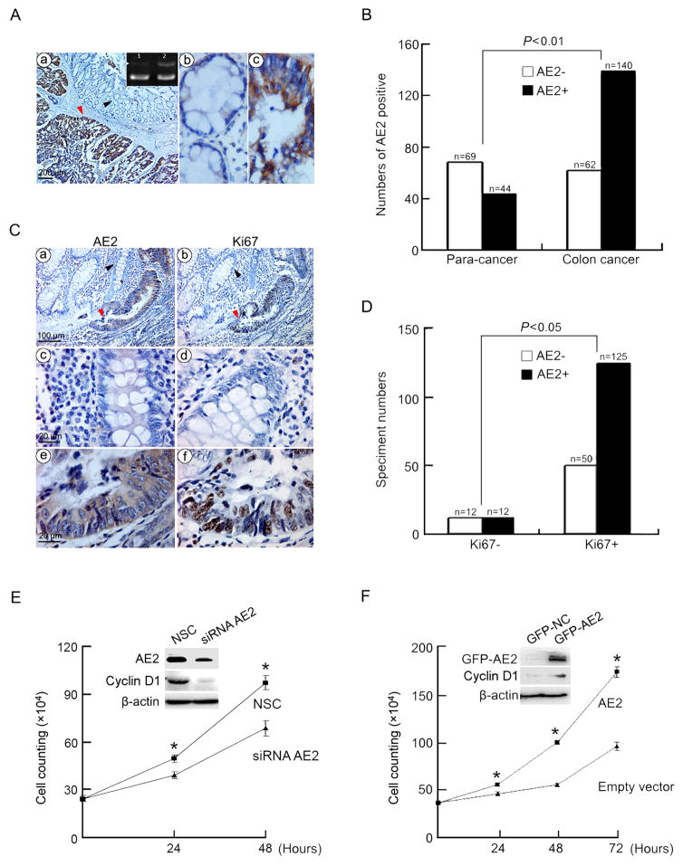
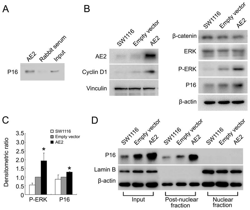
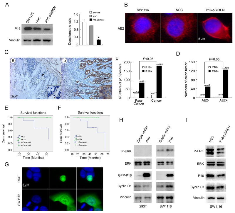
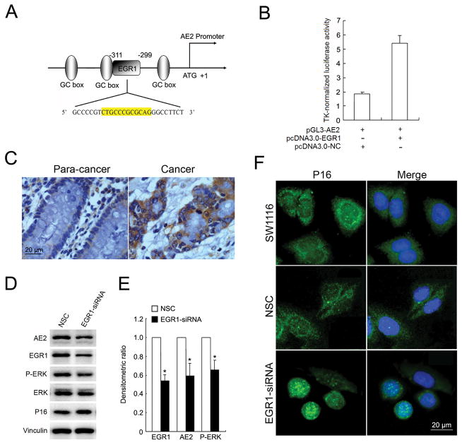
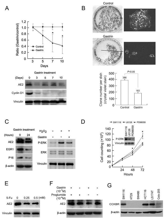
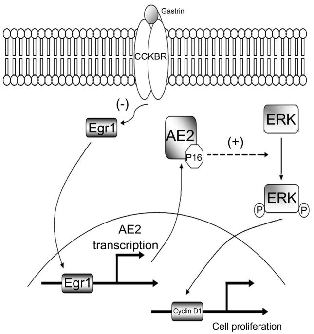
Human anion exchanger 2 (AE2) is a plasma membrane protein that regulates intracellular pH and cell volume. AE2 contributes to transepithelial transport of chloride and bicarbonate in normal colon and other epithelial tissues. We now report that AE2 overexpression in colon cancer cells is correlated with expression of the nuclear proliferation marker, Ki67. Survival analysis of 24 patients with colon cancer in early stage or 33 patients with tubular adenocarcinoma demonstrated that expression of AE2 is correlated with poor prognosis. Cellular and molecular experiments indicated that AE2 expression promoted proliferation of colon cancer cells. In addition, we found that transcription factor EGR1 underlies AE2 upregulation and the AE2 sequester p16INK4a (P16) in the cytoplasm of colon cancer cells. Cytoplasmic P16 enhanced ERK phosphorylation and promoted proliferation of colon cancer cells. Gastrin inhibited proliferation of colon cancer cells by suppressing expression of EGR1 and AE2 and by blocking ERK phosphorylation. Taken together, our data describe a novel EGR1/AE2/P16/P-ERK signaling pathway in colon carcinogenesis, with implications for pathologic prognosis and for novel therapeutic approaches.


Conflict of interest statement

Conflict-of-interest statement: The authors declare no conflict of interest.

Figures

Figure 1
Figure 1. Overexpression of AE2 was correlated with cell proliferation in colon cancer
(A) AE2 mRNA was analyzed by RT-PCR (a, upper right corner: 1, adjacent normal tissue; 2, colon cancer tissue; upper bands, AE2, lower bands, GAPDH). AE2 protein was analyzed by immunohistochemistry. The arrowed profiles in (a) were enlarged in (b, black arrow) and (c, red arrow). Original magnifications ×40 in a, x400 in b and c. (B) AE2 expression in para-cancer and colon cancer tissues was statistically analyzed. (C) Co-localization of AE2 and Ki67 in colon cancer tissues detected by immunohistochemistry. The arrowed profiles in (a) were enlarged in (c, black arrow) and (e, red arrow). The arrowed profiles in (b) were enlarged in (d, black arrow) and (f, red arrow). Original magnifications x200 in a and b, x1000 in c, d, e and f. (D) Association of histochemically detected expression of AE2 and Ki67 in colon cancer sections. (E) Growth curves of SW1116 cells transfected with siRNA-AE2 or siRNA-NC (NSC). Inset, immunoblot analysis of AE2 and cyclin D1. * P<0.05 (n=3). (F) Growth curves of SW1116 cells transfected with pEGFP-AE2 or pEGFP-C1 empty vector (control). Inset, immunoblot analysis of AE2 and cyclin D1. * P<0.05 (n=3).
Figure 2
Figure 2. Interaction of AE2 and P16 in cytoplasm was associated with elevated P-ERK abundance in colon cancer cells
(A) Interaction of AE2 with P16 in SW1116 cells. Cell lysates were subjected to immunoprecipitation with antibodies to AE2 or with rabbit serum. Immunoprecipitates were then immunoblotted with antibody to P16. (B) Immunoblot detection of cyclin D1, β-catenin, ERK, P-ERK and P16 proteins in SW1116 cells 24 hrs after transfection with pEGFP-AE2 or with pEGFP-C1 empty vector. (C) Densitometric scanning ratios of b-actin-normalized expression of P-ERK and P16 protein expression. Results were expressed as mean ± SD. *P<0.05 as compared with empty vector (n=3). (D) Nuclear and post-nuclear fractions of SW1116 cells were separated 24 hrs post-transfection with pEGFP-AE2 or pEGFP-C1 empty vector, then assessed for expression of P16 and Lamin B by immunoblot.
Figure 3
Figure 3. Coexpression of AE2 and P16 is correlated with poor prognosis of colon cancer patients through ERK pathway
(A) Down-regulation of P16 protein in SW1116 cells by pSIREN-P16 but not by pSIREN-NC vector (NSC). Densitometric P16 protein expression was normalized to vinculin expression. Results were expressed as mean ± SD. * P<0.05 as compared with NSC (n=3). (B) Immunofluorescence assay of AE2 (red) in SW1116 cells transfected with pSIREN-P16 or pSIREN-NC empty vector (NSC). Blue, DAPI-stained nuclei (original magnification ×400). (C) P16 expression was analyzed by immunohistochemistry in adjacent normal (a, para-cancer) and colon cancer tissues (b, original magnification ×100) and statistically analyzed (c). The arrowed regions were enlarged in lower right corner (original magnification ×1000). (D) Correlation analysis of AE2 and cytoplasmic P16 in colon cancer specimens. (E) Twenty-four patients were followed up for survival to assess AE2 expression as a prognostic factor. Kaplan-Meier survival curves and Log-rank test comparing patient groups with colon cancer segregated according to histologic detection of cytoplasmic AE2 expression. AE2-positive cases showed a significantly lower survival rate (63.6%) compared with AE2-negative patients (100%) (Log rank = 4.121, P= 0.042). (F) Thirty-three patients with tubular colon adenocarcinoma were followed up for survival to assess AE2 expression as a prognostic factor. Kaplan-Meier survival curves and Log-rank test comparing patient groups with colon cancer segregated according to histologic detection of cytoplasmic AE2 expression. AE2-positive cases showed a significantly lower survival rate (61.1%) compared with AE2-negative patients (100%) (Log rank = 4.699, P= 0.030). (G) Immunofluorescence assay of P16 (green) in HEK293T and SW1116 cells transfected with pEGFP-P16 vector. Blue, DAPI-stained nuclei (original magnification ×400). (H) Immunoblot detection of cyclin D1, ERK, P-ERK and P16 in HEK 293T and SW1116 cells transfected with pEGFP-P16 or pEGFP-C1 empty vector. (I) Immunoblot measurement of cyclin D1, ERK, P-ERK and P16 in SW1116 cells transfected with pSIREN-P16 or pSIREN-NC vector (NSC). pSIREN-NC is scrambled-sequence control.
Figure 4
Figure 4. AE2 expression is regulated by EGR1
(A) Schematic diagram of human AE2 proximal promoter reigion, containing three GC boxes and an EGR1 binding site. The numbers indicate the position relative to the ATG (+1). (B) SW1116 cells were transiently co-transfected with pGL3-AE2 luciferase reporter vector and pcDNA3.0-EGR1 or pcDNA3.0-NC vector (control). AE2 promoter activity was evaluated by luciferase assay. (C) Immunohistochemistry staining of EGR1 in colon cancer and para-cancer tissues (original magnification ×400). (D) Immunoblotting analysis of AE2, ERK, P-ERK and P16 expressions in SW1116 cells transfected with EGR1-siRNA or NC-siRNA (NSC). (E) Densitometric, vinculin-normalized relative expression of EGR1, AE2 and P-ERK proteins. Results are expressed as mean ± SD from three independent experiments. (F) Immunofluorescence assay of P16 (green) in SW1116 cells and cells transfected with EGR1-siRNA or NC-siRNA. Blue, DAPI-stained nuclei (original magnification ×400).
Figure 5
Figure 5. Gastrin suppresses growth and clone formation of SW1116 cells by inhibiting the EGR1/AE2/P16/P-ERK pathway
(A) SW1116 cell number was assessed following growth in the absence or presence of 10−7 M gastrin for periods of 3, 5, 7, and 10 days. Cell counts are expressed as values normalized to those of untreated cells (upper panel). * P<0.05 (n=3). Expression of AE2 and cyclin D1 was detected by immunoblot (lower panel). (B) Effect of 10 days’ treatment of SW1116 cells with 10−7M gastrin was assessed by clonogenic assay using crystal violet staining (upper panel, left) and phase contrast microscopy (upper panel, right). Number of clones per dish after 10 days in the absence or presence of 10−7M gastrin (lower panel), * P<0.05 (n=3). (C) Immunoblot analysis of AE2, EGR1, and P16 in SW1116 cells treated without or with 10−7M gastrin for 24 hrs (left panel). Immunoblot analysis of ERK and P-ERK in SW1116 cells treated with 100 μM H2O2 in the absence or presence of 10−7M gastrin for 24 hrs (right panel). (D) Cell growth of SW1116 cells treated with ERK inhibitors (30 μM U0126 or 50 μM PD98059). Inset, immunoblot analysis of P-ERK. * P<0.05 (n=3). (E) Immunoblot analysis of AE2 in SW1116 cells treated with 0.25 or 0.5 mM 5-Fluorouracil (5-Fu) for 24 hrs. (F) Immunoblot detection of AE2 expression in SW1116 cells treated with 10−7M gastrin in the absence or presence of the cholecystokinin B receptor (CCKBR) antagonist proglumide (10−4 M). (G) Immunoblot detection of CCKBR expression in colon cancer cell lines.
Figure 6
Figure 6. Diagram of a proposed novel EGR1/AE2/P16/P-ERK signal pathway in colon cancer cells
EGR1 transcriptionally upregulates AE2 expression. AE2 polypeptide interacts with and sequesters P16 in the cancer cell cytoplasm. Cytoplasmic P16 promotes phosphorylation of ERK, which can translocate into the nucleus, and activate cyclin D1 to promote cell proliferation. Gastrin acts through CCKBR at the plasma membrane to decrease EGR1 expression. Blockade of the EGR1/AE2/P16/P-ERK signaling pathway inhibits growth of colon cancer cells.

Similar articles

Cited by

References

    1. Saif MW, Chu E. Biology of colorectal cancer. Cancer J. 2010;16:196–201. doi: 10.1097/PPO.0b013e3181e076af00130404-201005000-00003. [pii] - DOI - PubMed
    1. Plotnikov A, Chuderland D, Karamansha Y, Livnah O, Seger R. Nuclear extracellular signal-regulated kinase 1 and 2 translocation is mediated by casein kinase 2 and accelerated by autophosphorylation. Mol Cell Biol. 2011;31:3515–3530. doi: 10.1128/MCB.05424-11. MCB.05424-11 [pii] - DOI - PMC - PubMed
    1. Kang YH, Yang IJ, Shin HM. Herbal formula HMC05 prevents human aortic smooth muscle cell migration and proliferation by inhibiting the ERK1/2 MAPK signaling cascade. J Nat Med. 2011 doi: 10.1007/s11418-011-0573-3. - DOI - PubMed
    1. She QB, Halilovic E, Ye Q, Zhen W, Shirasawa S, Sasazuki T, Solit DB, Rosen N. 4E-BP1 is a key effector of the oncogenic activation of the AKT and ERK signaling pathways that integrates their function in tumors. Cancer Cell. 2010;18:39–51. doi: 10.1016/j.ccr.2010.05.023. S1535-6108(10)00235-7 [pii] - DOI - PMC - PubMed
    1. Alper SL. Molecular physiology of SLC4 anion exchangers. Exp Physiol. 2006;91:153–161. doi: 10.1113/expphysiol.2005.031765. expphysiol.2005.031765 [pii] - DOI - PubMed

Publication types

MeSH terms

LinkOut - more resources