. 2012 Jan 24;109(4):1092-7.
doi: 10.1073/pnas.1112495109. Epub 2012 Jan 6.

Mitochondrial localization and structure-based phosphate activation mechanism of Glutaminase C with implications for cancer metabolism

Affiliations

Mitochondrial localization and structure-based phosphate activation mechanism of Glutaminase C with implications for cancer metabolism

Alexandre Cassago et al. Proc Natl Acad Sci U S A. .

Abstract

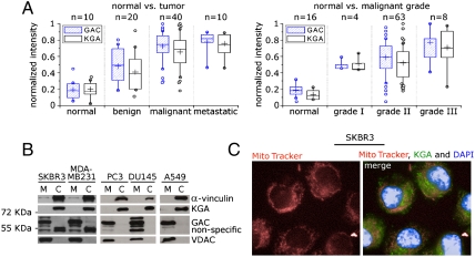
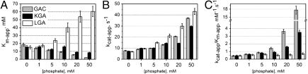
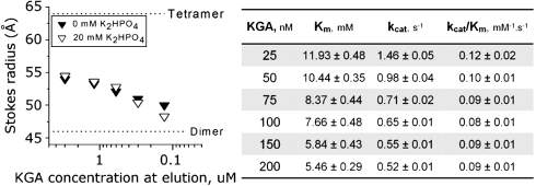
Glutamine is an essential nutrient for cancer cell proliferation, especially in the context of citric acid cycle anaplerosis. In this manuscript we present results that collectively demonstrate that, of the three major mammalian glutaminases identified to date, the lesser studied splice variant of the gene gls, known as Glutaminase C (GAC), is important for tumor metabolism. We show that, although levels of both the kidney-type isoforms are elevated in tumor vs. normal tissues, GAC is distinctly mitochondrial. GAC is also most responsive to the activator inorganic phosphate, the content of which is supposedly higher in mitochondria subject to hypoxia. Analysis of X-ray crystal structures of GAC in different bound states suggests a mechanism that introduces the tetramerization-induced lifting of a "gating loop" as essential for the phosphate-dependent activation process. Surprisingly, phosphate binds inside the catalytic pocket rather than at the oligomerization interface. Phosphate also mediates substrate entry by competing with glutamate. A greater tendency to oligomerize differentiates GAC from its alternatively spliced isoform and the cycling of phosphate in and out of the active site distinguishes it from the liver-type isozyme, which is known to be less dependent on this ion.


Conflict of interest statement

The authors declare no conflict of interest.

Figures

Fig. 1.
Fig. 1.
KGA and GAC are enhanced in cancer, but only GAC is found in mitochondria. (A) Box-and-whisker representation of the immunohystochemical analysis of human breast cancer tissue arrays. Isozyme-specific antibodies show that protein levels of both GAC and KGA increase in tumor tissues when compared to their normal healthy counterparts, and correlate both with the degree of malignancy (left box) and the grade of the tumor (right box). Intensities were normalized to the highest count measured. Lower and upper quartiles represent 25% and 75% of the data points and the middle band is the median. Whisker’s lengths define data between 10% and 90%. Outliers are shown as circles. Crosses indicate mean value of each set of data. (B) Fractioning of the breast SKBR3 and MDA-MB231, prostate PC3 and DU145 and lung A549 tumor cell lines followed by immunoblotting shows that KGA is found in the cytosol but not in the mitochondria, as opposed to GAC. (C) KGA cytoplasmic location was confirmed by immunofluorescence of SKBR3 cells stained with both MitoTracker and DAPI.
Fig. 2.
Fig. 2.
GAC has the highest activity in the presence of phosphate. (A) Kinetic analysis of the three isozymes shows that the apparent affinity for glutamine of KGA and GAC increases with higher concentrations of Pi, as opposed to what is observed for LGA, as suggested by their Km-app values. (B) Catalytic rates are increased, though at lower levels for KGA, as more phosphate is added to the reaction. (C) GAC becomes the most efficient isozyme already at concentrations of Pi around 10 mM.
Fig. 3.
Fig. 3.
The crystal structure of GAC. (A) Orthogonal views of the tetramer in cartoon representation. Circles indicate, in chain A, the two distinct domains comprising the full structure: amino-terminal and glutaminase. Though present in the crystallized construct, the C terminus is heterogeneous in conformation and could not be successfully modeled. Stereographic views of the cationic active site of GAC, represented by electrostatic surface mapping (formula image
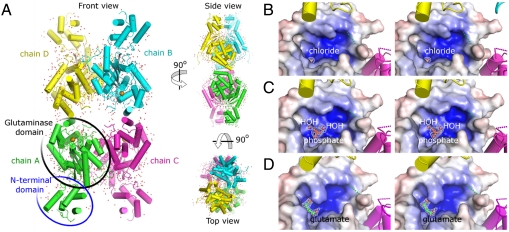
) for the three crystal forms, show the presence of chloride (B), phosphate (C) and L-glutamate (D). 2Fo-Fc Fourier electron density maps of the ligands are contoured at 1σ.
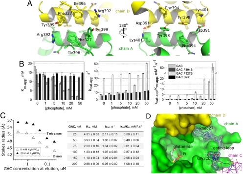
Fig. 4.
Fig. 4.
Activation mechanism based on the gating loop and inorganic phosphate. (A) Perpendicular views of the tetramer interface, defined by the symmetric stacking of the helix between residues Asp391 and Lys401 from each monomer. (B) Enzymatic characterization of point and deletion mutants in comparison to the wild-type enzyme show loss-of-function for GAC.F394S and GAC.DelC and gain-of-function for GAC.F327S. (C) Decrease in the Km for glutamine of GAC (right box) correlates with its protein concentration-dependent oligomerization profile, as determined by size-exclusion chromatography. Introduction of phosphate to the protein solution results in shifts of the equilibrium towards higher molecular weight species. (D) Top view of the active site of GAC and its proximity to the gating loop (dashed line between Gly320 and Phe327). Phe327 is part of the buried interface upon tetramer formation. Fourier 2Fo-Fc electron density map (contoured at 1σ) shows that residues flanking the loop are well ordered. Relative positions of Pi, L-glutamate and the chloride ion can be seen inside the active site, as a result of structure superposition.
Fig. 5.
Fig. 5.
Concentration-dependent oligomerization and activation profile of KGA. This isoform responds much more slowly than GAC to increases in protein concentration regarding shifts in the equilibrium towards species of higher Stokes radius. Furthermore, the addition of phosphate has no effects on the overall profile (left box). The slowed response is also reflected in less significant changes in the enzyme’s Km (right box), when compared to GAC (Fig. 4C).

Similar articles

Cited by

  • Two Faces of Glutaminase GLS2 in Carcinogenesis.
    Buczkowska J, Szeliga M. Buczkowska J, et al. Cancers (Basel). 2023 Nov 24;15(23):5566. doi: 10.3390/cancers15235566. Cancers (Basel). 2023. PMID: 38067269 Free PMC article. Review.
  • Structure and activation mechanism of the human liver-type glutaminase GLS2.
    Ferreira IM, Quesñay JEN, Bastos AC, Rodrigues CT, Vollmar M, Krojer T, Strain-Damerell C, Burgess-Brown NA, von Delft F, Yue WW, Dias SM, Ambrosio AL. Ferreira IM, et al. Biochimie. 2021 Jun;185:96-104. doi: 10.1016/j.biochi.2021.03.009. Epub 2021 Mar 18. Biochimie. 2021. PMID: 33746066 Free PMC article.
  • Decrypting noncoding RNA interactions, structures, and functional networks.
    Fabbri M, Girnita L, Varani G, Calin GA. Fabbri M, et al. Genome Res. 2019 Sep;29(9):1377-1388. doi: 10.1101/gr.247239.118. Epub 2019 Aug 21. Genome Res. 2019. PMID: 31434680 Free PMC article. Review.
  • MYCN mediates cysteine addiction and sensitizes neuroblastoma to ferroptosis.
    Alborzinia H, Flórez AF, Kreth S, Brückner LM, Yildiz U, Gartlgruber M, Odoni DI, Poschet G, Garbowicz K, Shao C, Klein C, Meier J, Zeisberger P, Nadler-Holly M, Ziehm M, Paul F, Burhenne J, Bell E, Shaikhkarami M, Würth R, Stainczyk SA, Wecht EM, Kreth J, Büttner M, Ishaque N, Schlesner M, Nicke B, Stresemann C, Llamazares-Prada M, Reiling JH, Fischer M, Amit I, Selbach M, Herrmann C, Wölfl S, Henrich KO, Höfer T, Trumpp A, Westermann F. Alborzinia H, et al. Nat Cancer. 2022 Apr;3(4):471-485. doi: 10.1038/s43018-022-00355-4. Epub 2022 Apr 28. Nat Cancer. 2022. PMID: 35484422 Free PMC article.
  • Oncometabolites-A Link between Cancer Cells and Tumor Microenvironment.
    Baryła M, Semeniuk-Wojtaś A, Róg L, Kraj L, Małyszko M, Stec R. Baryła M, et al. Biology (Basel). 2022 Feb 9;11(2):270. doi: 10.3390/biology11020270. Biology (Basel). 2022. PMID: 35205136 Free PMC article. Review.

References

    1. Warburg O. On the origin of cancer cells. Science. 1956;123:309–314. - PubMed
    1. Hsu PP, Sabattini DM. Cancer cell metabolism: Warburg and beyond. Cell. 2008;134:703–707. - PubMed
    1. Heiden MG, Cantley LC, Thompson CB. Understanding the Warburg Effect: the metabolic requirements of cell proliferation. Science. 2009;324:1029–1036. - PMC - PubMed
    1. DeBerardines RJ, Cheng T. Q’s next: the diverse functions of glutamine in metabolism, cell biology and cancer. Oncogene. 2010;29:313–324. - PMC - PubMed
    1. Aledo JC, Gómez-Fabre PM, Olalla L, Márquez J. Identification of two human glutaminase loci and tissue-specific expression of the two related genes. Mamm Genome. 2000;11:1107–1110. - PubMed

Publication types

Associated data