Review
. 2012 Jun;14(6):416-27.
doi: 10.1002/jgm.1648.

CD40-targeted adenoviral cancer vaccines: the long and winding road to the clinic

Affiliations
Review

CD40-targeted adenoviral cancer vaccines: the long and winding road to the clinic

Basav N Hangalapura et al. J Gene Med. 2012 Jun.

Abstract

The ability of dendritic cells (DCs) to orchestrate innate and adaptive immune responses has been exploited to develop potent anti-cancer immunotherapies. Recent clinical trials exploring the efficacy of ex vivo modified autologous DC-based vaccines have reported some promising results. However, in vitro generation of autologous DCs for clinical administration, their loading with tumor associated antigens (TAAs) and their activation, is laborious and expensive, and, as a result of inter-individual variability in the personalized vaccines, remains poorly standardized. An attractive alternative approach is to load resident DCs in vivo by targeted delivery of TAAs, using viral vectors and activating them simultaneously. To this end, we have constructed genetically-modified adenoviral (Ad) vectors and bispecific adaptor molecules to retarget Ad vectors encoding TAAs to the CD40 receptor on DCs. Pre-clinical human and murine studies conducted so far have clearly demonstrated the suitability of a 'two-component' (i.e. Ad and adaptor molecule) configuration for targeted modification of DCs in vivo for cancer immunotherapy. This review summarizes recent progress in the development of CD40-targeted Ad-based cancer vaccines and highlights pre-clinical issues in the clinical translation of this approach.


Figures

Figure 1
Figure 1
A schematic of Ad5 and its mode of infection of permissive target cells. Adenoviruses consist of an icosahedral capsid with protruding trimeric fibers at vertices. The fiber trimer comprises an N-terminal tail, a central shaft and a globular knob. Ad5 infects permissive cells through the fiber knob domain binding the primary docking receptor, the Coxsackie Adenovirus Receptor (CAR), followed by receptor mediated endocytosis, which involves RGD sequences in the Ad5 penton base binding to integrins on the target cell surface.
Figure 2
Figure 2
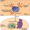
Schematic representation of the mode of action of CD40-targeted Ad-based vaccination. Both Langerhans Cells (LC) and dermal DC can be recruited to the dermis where CD40-targeted Ad5 vectors are delivered, leading to selective and high-efficiency in situ transduction of the dermally located, migratory DC. Roughly, three different CD40 targeting configurations can be distinguished: 1) a two-component configuration consisting of a bispecific adaptor fusion protein, binding the Ad5 fiber knob through soluble CAR (sCAR) or an anti-knob mAb; 2) A recombinant Ad5 with CD40L sequences incorporated into the fiber knob; 3) A recombinant mosaic Ad5 virus consisting of CAR-binding ablated wt fibers (ΔTAYT) and chimeric fibers incorporating CD40L sequences. The transduction and simultaneous activation of LC and dermal DC by CD40-targeted Ad, will result in their maturation and migration to skin draining lymph nodes, followed by presentation of endogenously processed transgenic tumor-associated antigens in the context of MHC molecules to T cells with all the necessary co-stimulatory signals to induce effective activation and expansion of tumor antigen-specific cytotoxic T cells in vivo.
Figure 3
Figure 3
Ex vivo human model systems for the assessment of enhanced and selective transduction of Dendritic cells (DC) by CD40-targeted Ad in human skin and skin draining lymph nodes (LN). A) Intradermal injection of Ad vectors into healthy human skin (obtained from cosmetic plastic surgery procedures). Note the typical formation of an intradermal urticus at the site of injection. Biopsies are taken of the injection sites (6mm in diameter; asterisks mark examples of post-biopsy scars with exposed dermis) and the explants are cultured floating in medium with the epidermal side up. After 2–3 days migrated DC are harvested for further analyses. B) The transduction rate of skin explant-emigrated DC was determined by flow cytometry. The number of migrated DC per explant was quantitated and the number of migrated transduced DC per explant calculated. Each data point in the graph represents an average of 10–20 explants per tested donor. Differences between untargeted Ad5 and CD40-targeted Ad5 were significant in a paired T test (*P<0.05)[50]. Insert shows fluorescence microscopic photograph (400×) of a transduced DC expressing GFP. C) Fluorescence-microscopic image (100×) shows GFP transgene expression in cells exhibiting DC morphology following transduction of SLN cells with CD40-Ad-GFP and lymphocyte clustering after CD40-mediated transduction, indicative of induced DC maturation and ongoing antigen presentation and T cell expansion. (D) A fluorescence-microscopic image (400×) shows GFP transgene expression in a cell exhibiting typical mature DC morphology following transduction of human SLN cells with CD40-Ad-GFP. Expansion of MART-126–35L-specific CD8+ T cell rates in an SLN suspension (derived from an early-stage melanoma patient) upon transduction with CD40-Ad-MART-1 (`pre” versus “post”)[51].
Figure 4
Figure 4
CD40-targeted Ad5 vector generation by genetic modification of its fiber. A) A schematic representation of the wild-type trimeric fiber structure of Ad5, comprising an N-terminal tail, a central shaft and a globular knob. B) The structure of bacteriophage T4 fibritin protein containing a central helical domain, a trimerization domain and a C-terminal 6-His motif. Trimeric CD40L was fused to the C-terminus of the artificial fiber. C) A chimeric CD40-targeted fiber protein comprising the Ad5 fiber-T4 fibritin-CD40 ligand. Boxes highlight the source of truncated parts of the wild-type fiber, bacteriophage T fibritin and CD40L used for generation of the chimeric fiber protein. The tail of the fiber anchors the fiber-fibritin-CD40L chimera in the Ad virion; a fragment of the fibritin protein provides trimerization of the molecule, while the CD40-ligand mediates binding to CD40 receptor.
Figure 5
Figure 5
Intradermal delivery of CD40-targeted Ad5 in BL6 mice leads to selective transduction of dendritic cells (DC) in the draining Lymph Node (LN). Intradermal injection of untargeted-Ad led to massive transduction of large CD11c macrophage-like cells, predominantly in the marginal zones of the draining Lymph Node (LN): A) immunofluorescence microscopic image of GFP expression, 100×; dotted line demarkates LN edge. Insert shows a bioluminescence pseudocolor heatmap image of levels of GFP expression in the draining LN (red: high, green: intermediate, and blue: low expression) at 24h post intradermal administration of Ad-GFP. B) GFP expression and CD11c staining (red) show the transduced cells to be CD11c. Cell nuclei were visualized using DAPI (blue), 400×. Intradermal injection of CD40-targeted Ad5 led to selective transduction of paracortical DC: C) Weak GFP expression was apparent in cells scattered throughout the LN (100×); dotted line demarkates LN edge. Insert shows a bioluminescence pseudocolor heatmap image of levels of GFP expression in the draining LN (blue: low expression) at 24h post intradermal administration of CD40-targeted Ad5-GFP. D) GFP expression and CD11c staining (red) show the transduced cells to be CD11c+ DC. Cell nuclei were visualized using DAPI (blue), 400×. Arrows denote CD11c+ GFP-expressing DC.
Figure 6
Figure 6
The long and winding road from the lab to the clinic: a timeline of highlights in the generation and pre-clinical evaluation of a CD40-targeted adenoviral cancer vaccine. 1999: A bispecific antibody conjugate was generated through chemical conjugation of a Fab fragment of a neutralizing anti-fiber-knob monoclonal antibody (mAb) to an agonistic anti-CD40 mAb. Efficient human monocyte-derived DC transduction and induction of maturation by CD40-targeted Ad was demonstrated using this bispecific Ab conjugate[48]. 2000: Using a similar mCD40-targeting chemical conjugate, improved efficacy of an ex vivo generated Bone Marrow-derived DC vaccine against Human Papillomavirus-induced tumor cells in a murine model was demonstrated[54]. 2002: The ability of CD40-targeted Ad to selectively transduce and activate dermally located DC in situ while ensuring their migration and enhancing their antigen specific CD8+ T cell priming ability, was demonstrated using a human skin explant model[50]. 2003: Second generation CD40-targeting conjugates for Ad5 consisting of an anti-CD40 single chain Fv fragment fused to either soluble CAR or an anti-Ad5 fiber knob scFv, were developed. The ability of such a second generation CD40-targeting conjugate to enhance gene transfer to DC in vitro and improve their CD8+ T cell activation capacity was demonstrated[55, 56]. 2005: A genetically targeted mosaic Ad vector directed to CD40-expressing cells was generated and its efficiency in transduction and activation of DC in human skin explants was demonstrated[49, 60]. 2006: An effort to translate this configuration to the clinic as a CEA-encoding colon cancer vaccine came to a halt when the NIH Biological Resources Branch Oversight Committee deemed the production process and the composition of the resulting mosaic fiber Ad vectors too uncontrolled and unpredictable. 2008: Construction and in vivo biodistribution analysis of a third generation adaptor fusion protein consisting of sCAR and the trimerized TNF-like active domain of CD40L[65]. 2009: The efficacy of a recombinant CD40-targeted Ad vaccine to induce antigen-specific T cell and antibody responses was studied in a canine model[66]. 2010: Using a third generation sCAR-CD40L adaptor protein, selective transduction of mature DC in human lymph nodes and superior induction of high-avidity melanoma-reactive cytotoxic T cells was demonstrated[51]. 2011: Demonstration of in vivo targeting of DC in skin and LN and induction of efficacious anti-tumor immunity in a therapeutic setting, again using a third generation sCAR-CD40L adaptor in the B16 mouse melanoma model[67].

Comment in

References

    1. Shortman K, Lahoud MH. Caminschi I Improving vaccines by targeting antigens to dendritic cells. Exp Mol Med. 2009;41:61–66. - PMC - PubMed
    1. Tacken PJ, de Vries IJ, Torensma R, et al. Dendritic-cell immunotherapy: from ex vivo loading to in vivo targeting. Nat Rev Immunol. 2007;7:790–802. - PubMed
    1. Wei H, Wang S, Zhang D, et al. Targeted delivery of tumor antigens to activated dendritic cells via CD11c molecules induces potent antitumor immunity in mice. Clin Cancer Res. 2009;15:4612–4621. - PubMed
    1. Dickgreber N, Stoitzner P, Bai Y, et al. Targeting antigen to MHC class II molecules promotes efficient cross-presentation and enhances immunotherapy. J Immunol. 2009;182:1260–1269. - PubMed
    1. Tagliani E, Guermonprez P, Sepulveda J, et al. Selection of an antibody library identifies a pathway to induce immunity by targeting CD36 on steady-state CD8 alpha+ dendritic cells. J Immunol. 2008;180:3201–3209. - PubMed

Publication types